
In the medical field, bone defects from infections are a big risk. They can lead to osteonecrosis, which can harm patient outcomes and lengthen hospital stays. Consequently, there is a pressing demand for a bone graft substitute that effectively combines broad-spectrum antibacterial properties with osteoinductive capabilities.
Recently, Professor Guomin Wu and his team at Jilin University published a significant study in Bioactive Materials. The researchers titled the study "3D-printed zinc oxide nanoparticles modified barium titanate/hydroxyapatite ultrasound-responsive piezoelectric ceramic composite scaffold for treating infected bone defects."
They used digital light processing (DLP) 3D printing to create a piezoelectric ceramic scaffold. This scaffold includes zinc oxide nanoparticles (ZnO-NPs), modified barium titanate (BT), and hydroxyapatite (HA). This work offers a new way to treat infected bone defects.
Research Background and Challenges
The treatment of infected bone defects has two main challenges. First, bone graft materials often lack antibacterial properties. Second, these materials have limited ability to promote bone growth. Traditional bone graft materials often fail to meet both requirements.
Professor Wu's research focuses on solving these problems. He introduces ZnO-NPs to improve the piezoelectric and antibacterial properties of BT/HA composites. They keep their biocompatibility and osteoinductive features intact while doing this.
Research Methods and Innovations
The research team utilized DLP 3D printing technology to precisely construct ZnO-NPs-modified BT/HA piezoelectric ceramic scaffolds. Through in vitro experiments, the team validated the scaffold's antibacterial and osteogenic capabilities.
The results showed that adding ZnO-NPs greatly improved the piezoelectric properties of BT. It also increased the HA content in the ceramic scaffold system, which is important for bone regeneration. Furthermore, the combination of low-intensity pulsed ultrasound (LIPUS) and piezoelectric effects demonstrated the scaffold's remarkable osteogenic and angiogenic potential.
Experimental Results and Data Analysis
1.Antibacterial Performance: Lab tests showed that the ZnO-NPs-modified BT/HA scaffold had strong antibacterial effects. It worked well against Staphylococcus aureus and Escherichia coli. Plate counting and live/dead bacterial staining confirmed the scaffold's antibacterial efficacy.
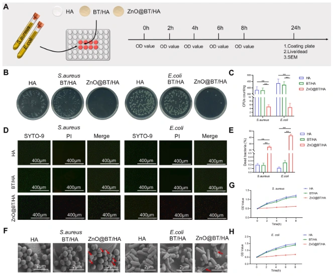
Figure 1: Results of antibacterial experiments showing the scaffold's effectiveness against bacteria
2.Biosafety: CCK-8 assays and live/dead cell staining demonstrated that the ZnO-NPs-modified scaffold had excellent biocompatibility, promoting cell proliferation and survival.
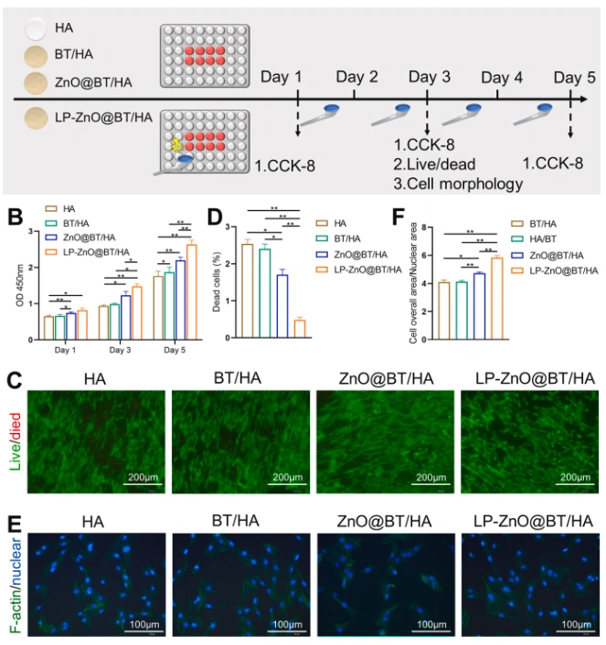
Figure 2: Biosafety test results confirming the scaffold's biocompatibility
3.Osteogenesis and Angiogenesis
ALP staining and immunofluorescence staining were used in the study. The results showed that the scaffold modified with ZnO-NPs significantly increased the levels of osteogenic proteins.
These proteins include OCN and OPN. It also increased angiogenic proteins, such as VEGF and HIF-1α. LIPUS stimulation further enhanced the scaffold's osteogenic and angiogenic capabilities.
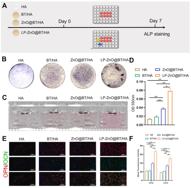
Figure 3: Results of osteogenic properties tests demonstrating the scaffold's bone regeneration potential
4.Transcriptomics analysis showed that the early growth response-1 (EGR1) gene is important for the scaffold's bone growth process. KEGG enrichment analysis further highlighted the importance of the "Parathyroid Hormone Synthesis, Secretion, and Action" signaling pathway in osteogenic differentiation.
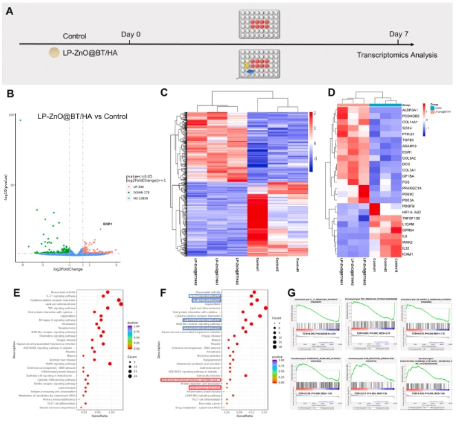
Figure 6: Transcriptomics analysis results identifying key genes and pathways involved in osteogenesis
Conclusion and Future Prospects
The study created a new 3D-printed piezoelectric ceramic scaffold. This scaffold combines the antibacterial properties of ZnO-NPs with the bone-promoting features of BT/HA.
The combined effects of LIPUS and piezoelectricity showed strong potential for bone growth and new blood vessel formation. This offers a hopeful approach for treating infected bone defects. In the future, this scaffold will be important in clinical use. It will help provide better treatment results for patients.