
Type 2 diabetes is a bone disease as much as a metabolic one. Patients fracture more easily, heal more slowly, and a defect that would close in a healthy patient can stay open for months. The clinical fallback — take bone from somewhere else and graft it in — is markedly less reliable in this group, and the usual explanations ("poor blood supply", "chronic inflammation") never told a surgeon what to change about the implant.
A 2026 study in Journal of Translational Medicine, from the Second Affiliated Hospital of Harbin Medical University with Tsinghua's biomedical engineering department and Dalian University of Technology, went after that question with an unusually complete chain: animal model first, multi-omics second, mechanism third, and only then a scaffold designed to hit the mechanism from two sides at once.
The conclusion is compact. Diabetes breaks bone healing at two independent points — a stalled step in glycolysis, and a blunted sense of mechanical load — and a scaffold that fixes only one of them leaves the cell adipogenic.
1. The gold standard fails first
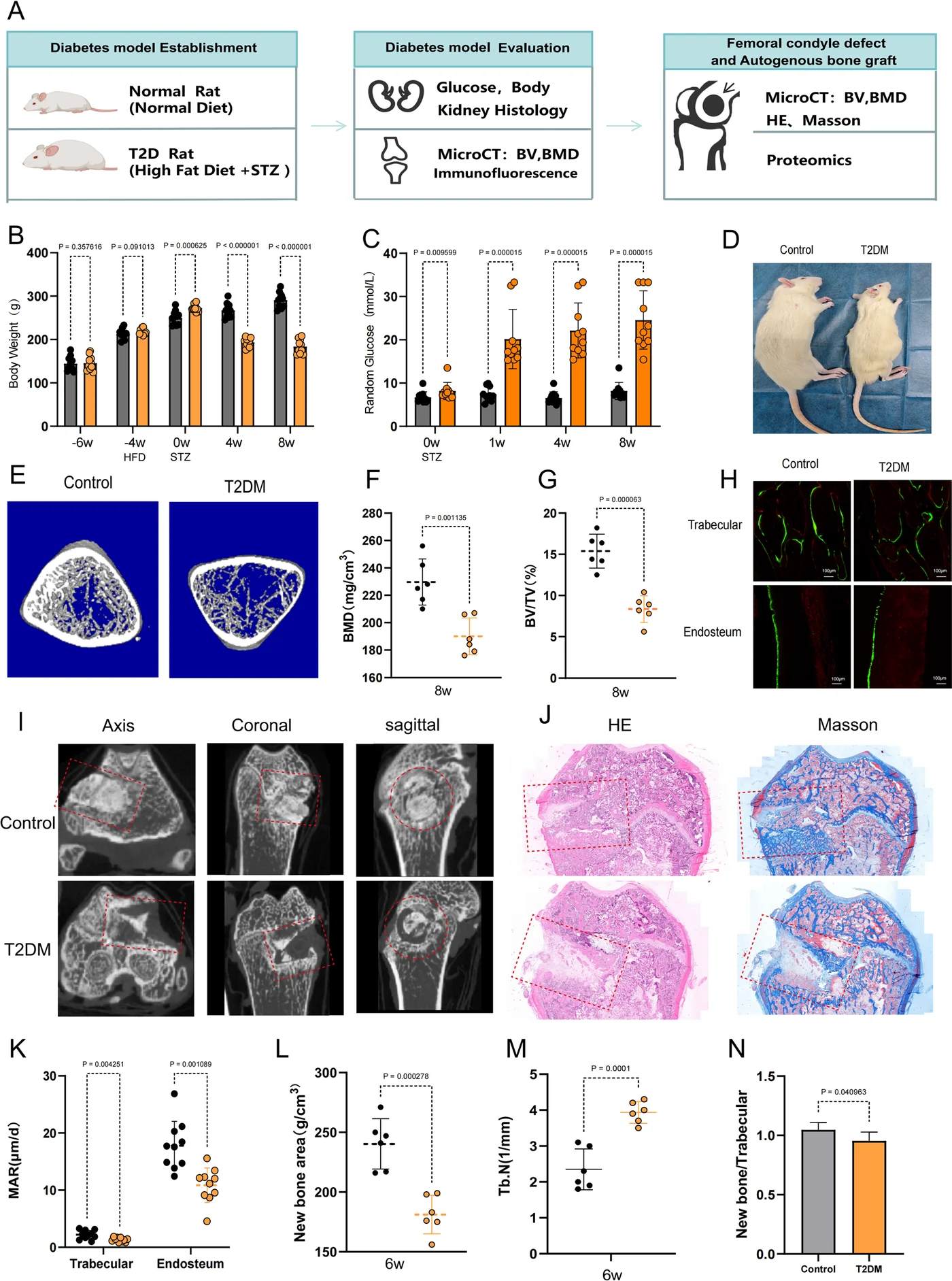
Male SD rats were put on a high-fat, high-sucrose diet for four weeks and then given streptozotocin at 35 mg/kg. Random blood glucose had to exceed 16.7 mmol/L, with glucose and insulin tolerance tests confirming insulin resistance; surgery followed after four more weeks of stable hyperglycaemia. A trephine created a 3 mm diameter × 4 mm deep cylindrical defect in the medial femoral condyle, filled with the animal's own cancellous bone — the clinical gold standard, in the same animal.
Six weeks later, micro-CT and HE/Masson histology showed the diabetic animals had significantly less new bone and fewer trabeculae. The study also normalised the result in a way worth stealing: it divided the mineral density of the newly formed bone by that of the native femoral bone. In control animals the ratio sat above 1; in diabetic animals it fell below 1. Same material, same surgeon, same defect — the graft was not the problem. The host was.
2. Two failures inside one cell
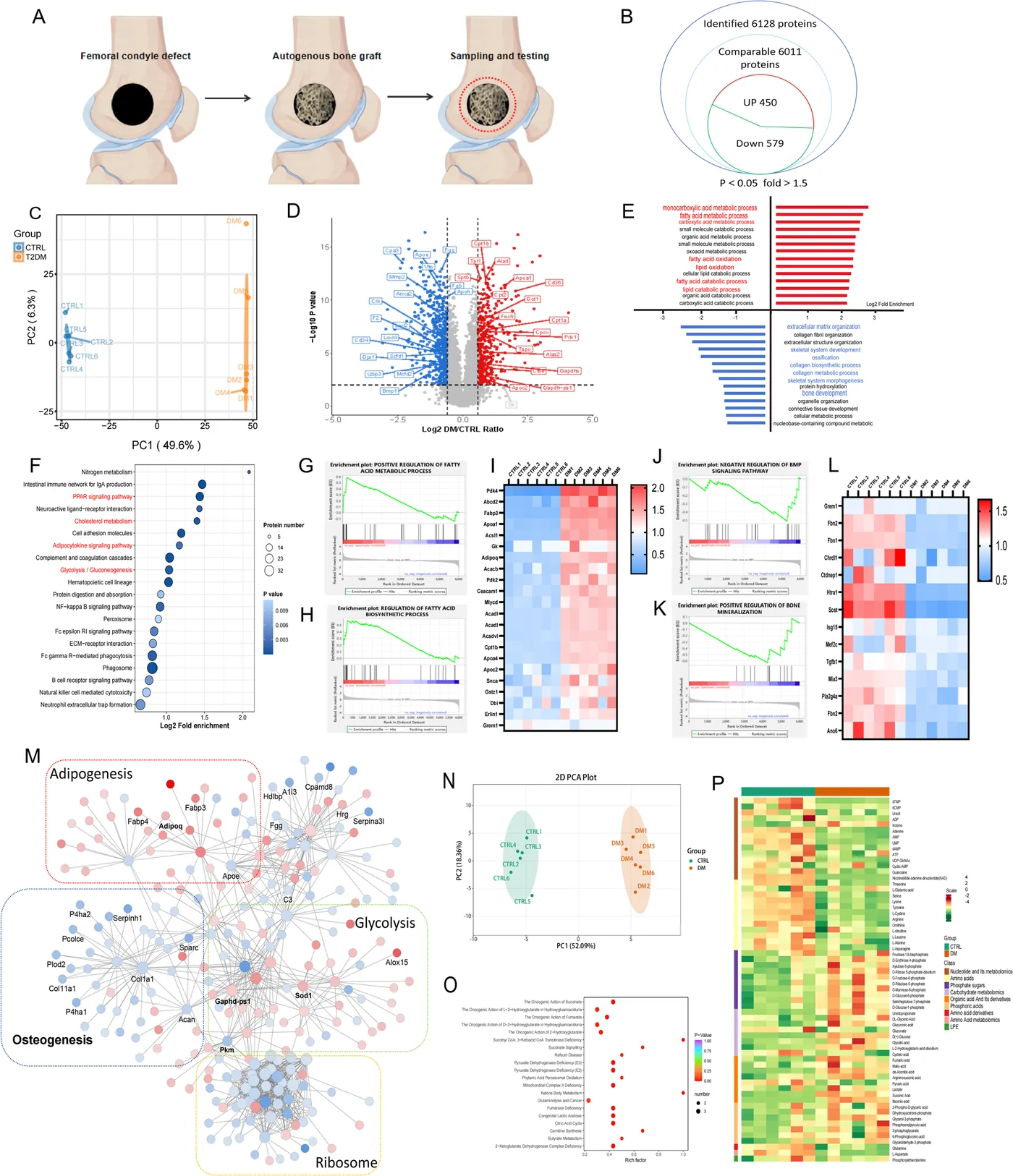
Proteomic sequencing of the healing region identified 6,128 proteins and 1,029 differentially expressed ones (450 up, 579 down, p < 0.05, fold change > 1.5). Gene set enrichment analysis split cleanly along fate: fatty acid synthesis and metabolism pathways were activated, BMP signalling and bone mineralisation were suppressed. A protein–protein interaction network collapsed the data into four clusters — osteogenesis, adipogenesis, glycolysis and ribosomes.
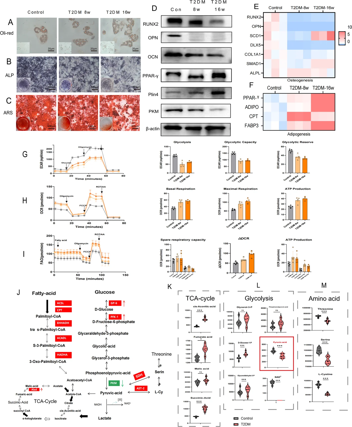
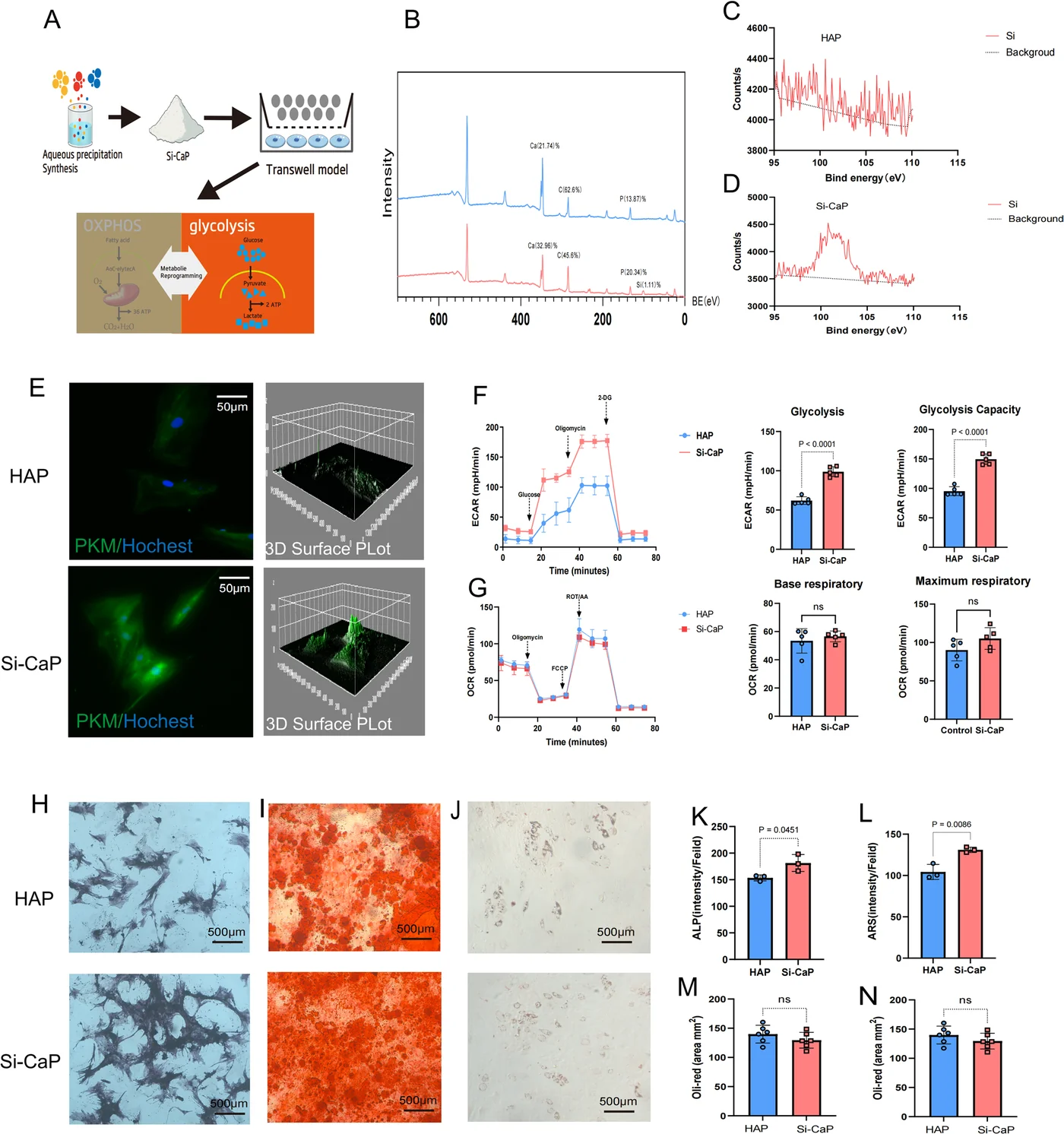
Targeted energy metabolomics pointed the same way, enriching in pyruvate metabolism, serine metabolism and the TCA cycle: glucose-related metabolites were elevated, amino acid metabolites were depleted. And when BMSCs were isolated at 8 and 16 weeks of diabetes, the phenotype deepened with time — more lipid droplets, weaker ALP staining, fewer mineralised nodules — while the Seahorse profile matched the tissue data: glycolytic capacity down, oxidative phosphorylation and fatty acid oxidation up as compensation.
Adipogenesis up, osteogenesis down, energy metabolism rewired. That is the phenotype. The interesting part is where it originates.
3. The bottleneck is PKM
Working backwards through glycolysis, the intermediates from glucose to phosphoenolpyruvate (PEP) and their enzymes were all higher in the diabetic group. Then the chain breaks: pyruvate kinase M (PKM), the enzyme that converts PEP to pyruvate, was down — confirmed at protein level in both the 8-week and 16-week groups — and its products, pyruvate and lactate, fell with it.
The authors call the result "somewhat contradictory": glycolytic metabolites are elevated, yet glycolytic flux is impaired. Their own data resolve it — every intermediate from glucose to PEP is up while PKM, the next step, is down, so intermediates accumulate behind the block and the flux measured downstream of it falls. The bottleneck is a single enzyme, which makes it a target: applying the PKM activator TEPP-46 reversed the defect, restoring pyruvate and lactate and reactivating glycolysis.
4. The cell stops feeling load
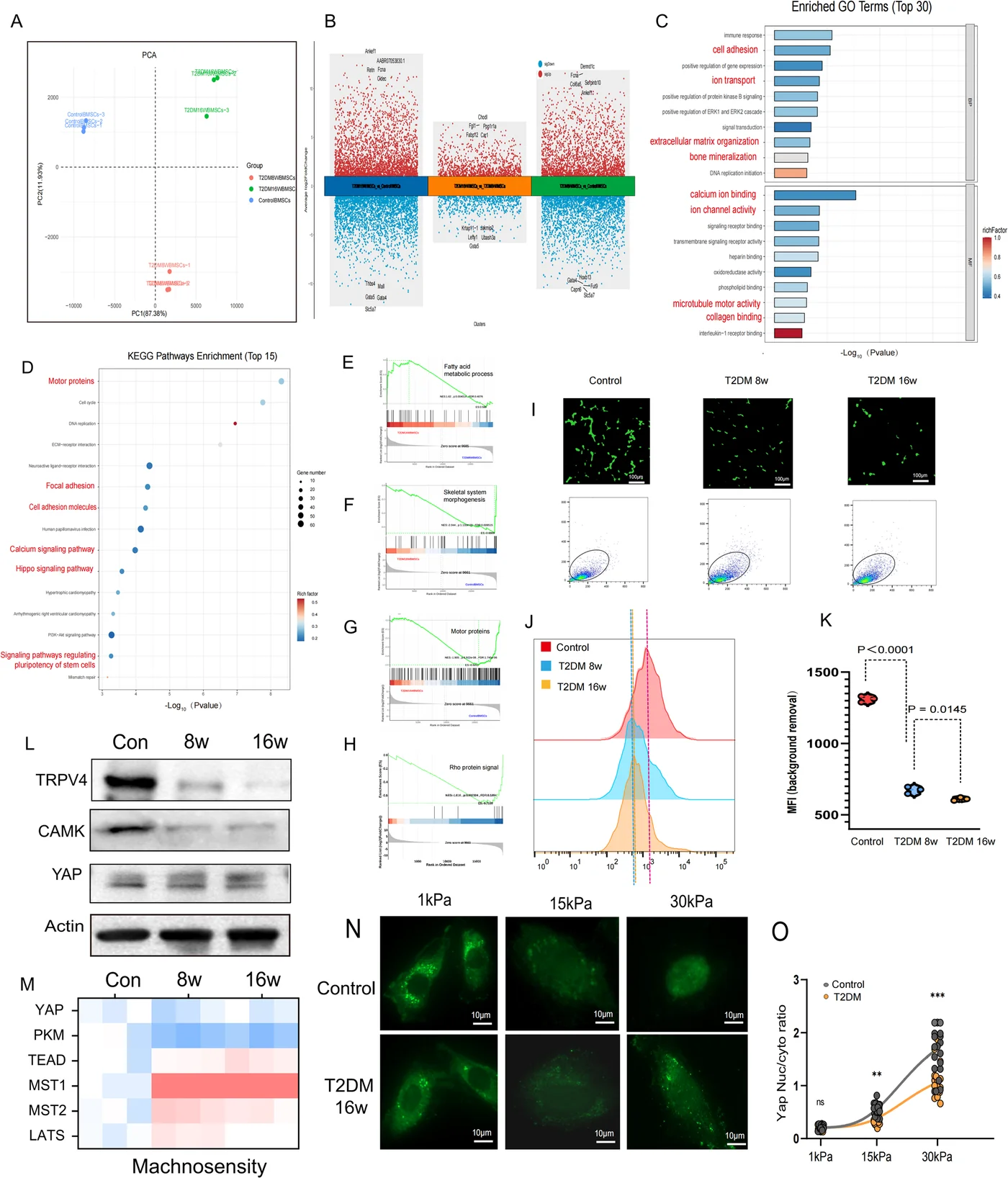
RNA sequencing of BMSCs from the 16-week group against controls returned 2,932 upregulated and 2,840 downregulated genes, enriched in motor proteins, calcium influx and Hippo signalling — the machinery a cell uses to feel its substrate. The calcium data contain the most telling detail in the paper. Using a calcium probe and flow cytometry, the proportion of cells showing calcium influx did not change. What collapsed was the peak influx per cell. Not a dead population — a desensitised one.
Two proteins behind that signal were down, the channel TRPV4 and calmodulin (CAMK); the kinases that phosphorylate and inactivate YAP, LATS and MST, were up. Cultured on hydrogels of 1, 15 and 30 kPa, diabetic BMSCs showed markedly less YAP nuclear localisation on the 15 and 30 kPa substrates, while total YAP was unchanged — a localisation defect, not an abundance defect.
That matters because YAP is the switch between the two fates. Nuclear YAP drives RUNX2 and osteogenesis; cytoplasmic YAP leaves PPARγ free to drive adipogenesis. A cell that cannot feel a stiff substrate behaves as if it were on a soft one — and becomes a fat cell.
5. Fixing the chemistry half: Si-CaP
Si-CaP is a silicon-substituted hydroxyapatite that releases Si ions; it was synthesised according to previously reported methods, and XPS showed a silicon peak between 110 and 95 eV. XRD matched the Si-CaP reference pattern, and FT-IR confirmed Si–O–Si bonding through a broadened 1050 cm⁻¹ band and a new feature at 750 cm⁻¹. In Tris-HCl the material released silicon ions continuously and stably over 24 hours, and CCK-8 showed no proliferation penalty on BMSCs even at three times that concentration.
In a Transwell co-culture, Si-CaP raised PKM expression and restored the glycolytic profile of diabetic BMSCs: basal and maximal glycolytic capacity, ATP production and glycolytic reserve all improved, while oxygen consumption rate did not change. Downstream, ALP rose at 7 days and calcium nodules at 21 days.
And then the honest result: Oil Red O staining showed no reduction in lipid droplet formation. Chemistry alone fixed the metabolism and promoted osteogenesis, but it did not switch off the adipogenic programme. The cells still had a choice, and they still leaned fat.
6. Fixing the geometry half: TPMS
So the authors changed the architecture. Two scaffolds were built with the same Si-CaP composition, the same 3 mm diameter × 4 mm height and the same porosity — one an orthogonal array of cylinders, the other a triply periodic minimal surface (TPMS) structure — and both were printed from Si-CaP by DLP. Nearly everything that follows is attributable to geometry alone.
On the cylinder scaffold the diabetic BMSCs spread nearly round. On the TPMS scaffold the concave surfaces forced them to elongate, stretching the actin cytoskeleton. Finite element analysis showed a different stress field in the TPMS pores; the paper's text calls it more concentrated and its figure caption calls it more uniform, which is best read together — the field is redistributed more evenly overall while local curvature raises the peak a cell actually feels. Compression testing agreed that the TPMS structure carried load better. Water contact angle was identical between the two, ruling out surface chemistry as the explanation.
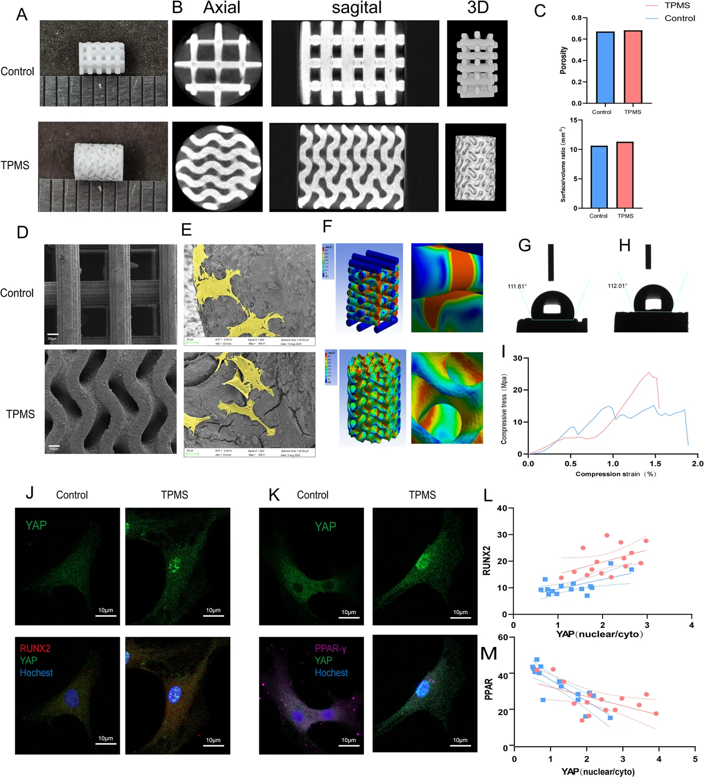
The result was visible in the nucleus. On the TPMS scaffold, a significantly larger fraction of YAP sat inside the nucleus, RUNX2 was up and PPAR was down. Across cells, the nuclear-to-cytoplasmic YAP ratio correlated positively with RUNX2 and negatively with PPAR. Then the causal check: cytochalasin D, which destroys actin filaments, scattered the cytoskeleton and markedly inhibited YAP nuclear translocation. The geometry-to-fate route runs through the cytoskeleton, or it does not run at all.
7. In vivo: the structured scaffold
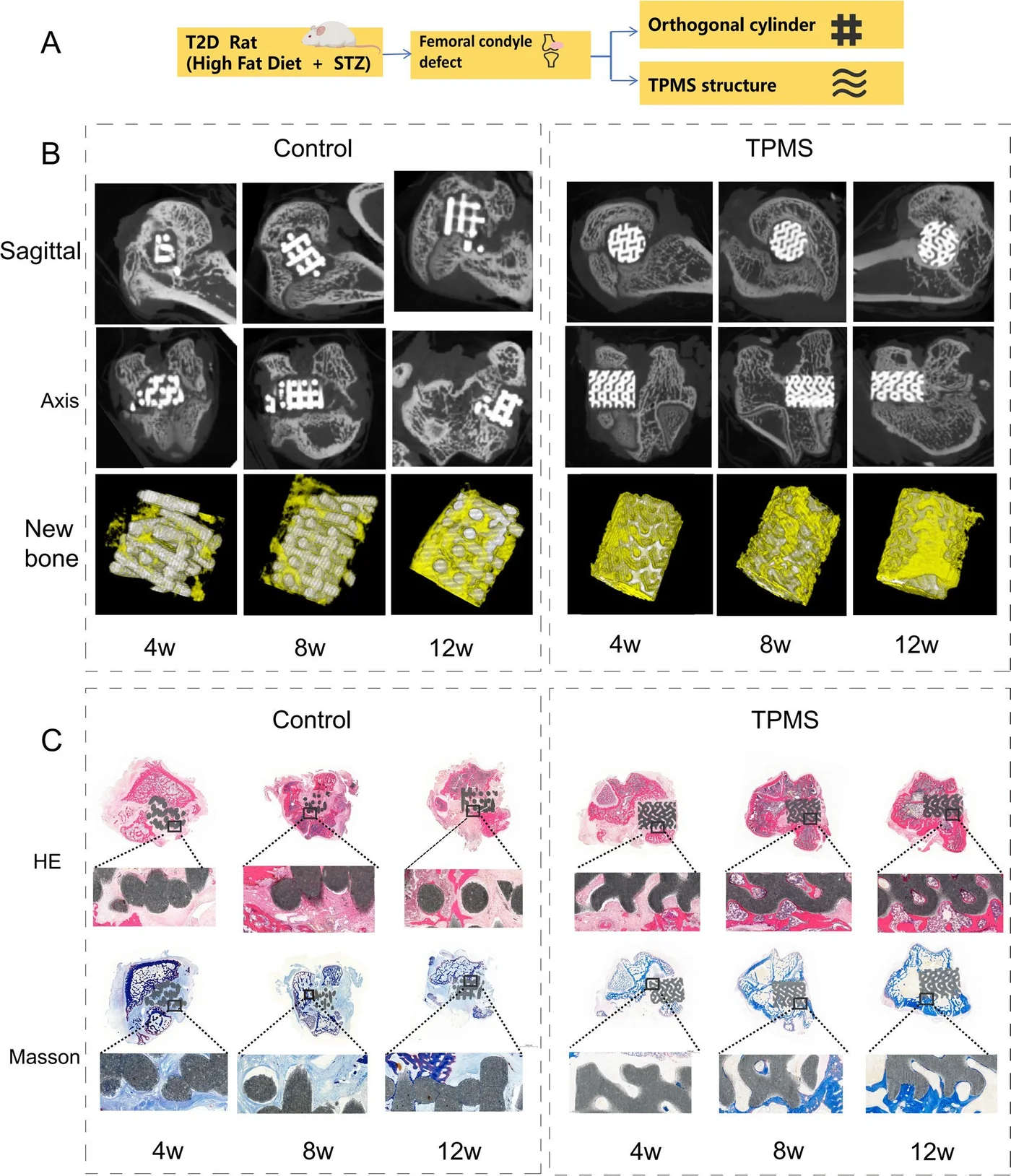
Diabetic rats received the 3 mm diameter × 4 mm deep femoral condyle defect eight weeks after diabetes induction, comparing the orthogonal cylinder scaffold against the TPMS scaffold. At 4, 8 and 12 weeks, micro-CT showed bone growing into the TPMS scaffold and new bone on its surface; bone mineral density, BV/TV and trabecular number were all higher than in the control group, and HE/Masson staining showed trabeculae connected to the scaffold with stronger collagen staining.
The study therefore closes the loop in a living diabetic animal, not only in a dish. What it does not close is the synergy claim — see the next section.
8. What the study does not show
- The in vivo experiment isolates architecture, not synergy. Both implanted groups are Si-CaP; there is no TPMS-HAP arm. The animal data therefore prove that topology helps, while the material's contribution rests on in vitro evidence. "Composition plus structure" is a reasonable synthesis of the whole paper, but the two were never tested apart in vivo.
- Osteoclasts were not examined. Bone healing is formation and resorption; this study measured only the formation side.
- No immunomodulation data. The authors state plainly that how Si-CaP composition and TPMS topology each shape the inflammatory cascade remains unknown.
- No long-term degradation or clinical comparison. Twelve weeks is the endpoint, and the scaffold was never compared head-to-head with a mainstream clinical bone graft.
- YAP's upstream trigger is a hypothesis. Whether the metabolic state changes YAP through glycosylation, phosphorylation or another post-translational modification was not tested; nor was whether silicon ions act on the cytoskeleton directly through integrins or RHO GTPase.
- Geometry is not a single variable. Curvature, pore shape and the local surface topography the cells sit on all change together between the two designs; the paper treats them as one "structure" factor and does not separate which of them drives YAP.
- Sample sizes are conventional but small. Animal groups run at n = 6 and RT-qPCR at n = 3, with larger n only where imaging was quantified; the Western blots carry no stated n.
9. What it means for ceramic 3D printing
Two things survive the caveats. First, architecture is a biological variable, not packaging. The same powder, the same porosity and the same outer dimensions produced a different cell fate because the pore walls curved — which means the design file is doing pharmacology, and it deserves the same rigour as the formulation.
Second, the two halves have to be designed together. Silicon substitution fixed the metabolism and left adipogenesis untouched; TPMS fixed the fate switch and would have had no glycolysis to work with. A scaffold that only does one is a half-treatment, and the paper is unusually clear about that.
Both halves are DLP problems. Printing Si-CaP into a defined pore architecture — 3 mm diameter, 4 mm tall, porosity held constant — is exactly what a ceramic vat-photopolymerisation system is built for; in this study, an ADT Ceram-D-L96 system. For bioceramic teams, the practical lesson is that a printer's value in bone work is not only feature resolution but repeatability of the pore geometry, because that geometry is now part of the therapy.
Source: Mo Y, Tang L, Zhang J, He W, Chen J, Zhang S, Chai B, Tian X, Yang Z, Zhang Y, Li P, Wang X. Decreased mechanosensitivity and metabolic reprogramming impair bone regeneration in type II diabetes: core mechanism and targeted scaffold design. Journal of Translational Medicine (2026). doi:10.1186/s12967-026-08925-1. Figures 1–7 are reproduced from the cited open-access article (CC BY 4.0) for discussion.