
Hydroxyapatite (HA) is a material widely used in bone tissue engineering, but its inherent brittleness and processing difficulty limit its application potential in porous bone stents. Recently, Professor Guo Wang of Guangxi University and Professor Long Yu's team published a research titled "Ionic substitution through bredigite doping for microstructure and performance adjustment in DLP 3D-printed TPMS porous HA bone scaffolds" in "Virtual and Physical Prototyping". They Hydroxyapatite (HA, chemical formula Ca₁₀(PO₄)₆(OH)₂) is modified by doping of brazilite (Ca₇MgSi₄O₁₆) to improve its mechanical and biological properties.
Original link: https://doi.org/10.1080/17452759.2024.2423840
Adventure Technology official website: https://www.adventuretech.cn
Research content
Hydroxyapatite is a typical calcium phosphate ceramic that is widely used in bone stent development because it is a major inorganic component of natural bone. However, HA has a high calcium-phosphorus ratio and strong chemical stability, and slow degradation rate, resulting in insufficient release of inorganic ions and limited biomineralization capacity, making it difficult to trigger sufficient biological responses.
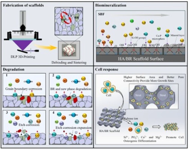
In this study, the ionic substitution modification of the hydroxyapatite (HA) bone scaffold was achieved through branlin doping, and the TPMS porous structure was constructed in combination with DLP 3D printing technology. The study found that doping significantly improves the microstructure and mechanical properties of the material, while enhancing biocompatibility and degradation behavior. The compressive strength of the scaffold reached the biomechanical standard after doping, and cell experiments verified its good osteocyte adhesion and proliferation performance .
The following are the research methods and data of the article:
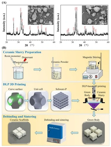
△Fig. 1, (A) XRD and SEM characterization of original HA and BR powders; (B) Schematic diagram of ceramic scaffolds prepared by DLP.
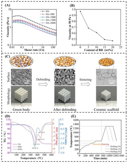
△Fig. 2, Rheological behavior of HA and HA/BR ceramic slurries and degreasing-sintering process of HA green body. (A) The relationship between viscosity and shear rate; (B) The relationship between viscosity and BR content when the shear rate is 30 s −1. (C) SEM surface micromorphology of HA scaffolds after DLP 3D printing, degreasing and sintering; and schematic diagrams of degreasing and sintering. (D) TG-DSC analysis of HA green body; (E) Designed degreasing and sintering process curves.
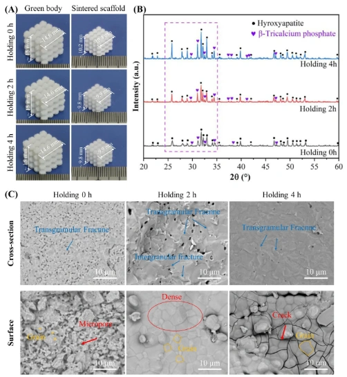
△Figure 3, Characterization of morphology, phase composition and microstructure of HA stents with different holding times. (A) Macro-morphology; (B) XRD pattern; (C) SEM morphology of cross-section and surface.
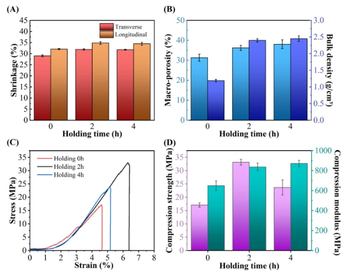
△Figure 4. Effect of insulation time on HA stent shrinkage, large porosity, bulk density and mechanical properties. (A) Transverse and longitudinal shrinkage; (B) Large porosity and bulk density; (C) Compression stress-strain curve; (D) Strength and modulus.
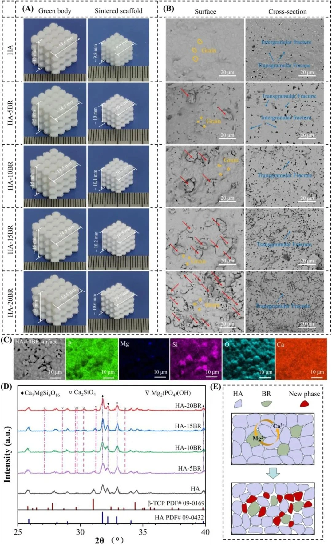
Figure 5. Topography, microstructure and phase composition of HA/BR composite scaffold. (A) Macromorphology; (B) Surface and cross-sectional morphology observed by SEM; (C) Elemental analysis of the surface by EDS mapping. (D) Phase analysis by XRD; (E) Schematic diagram showing ionic substitution between HA and BR to produce a new phase.
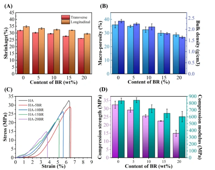
△Fig. 6, Effect of BR content on the shrinkage, large porosity, bulk density and mechanical properties of HA/BR stents. (A) Transverse and longitudinal shrinkage; (B) Large porosity and bulk density; (C) Compression stress-strain curve; (D) Strength and modulus.
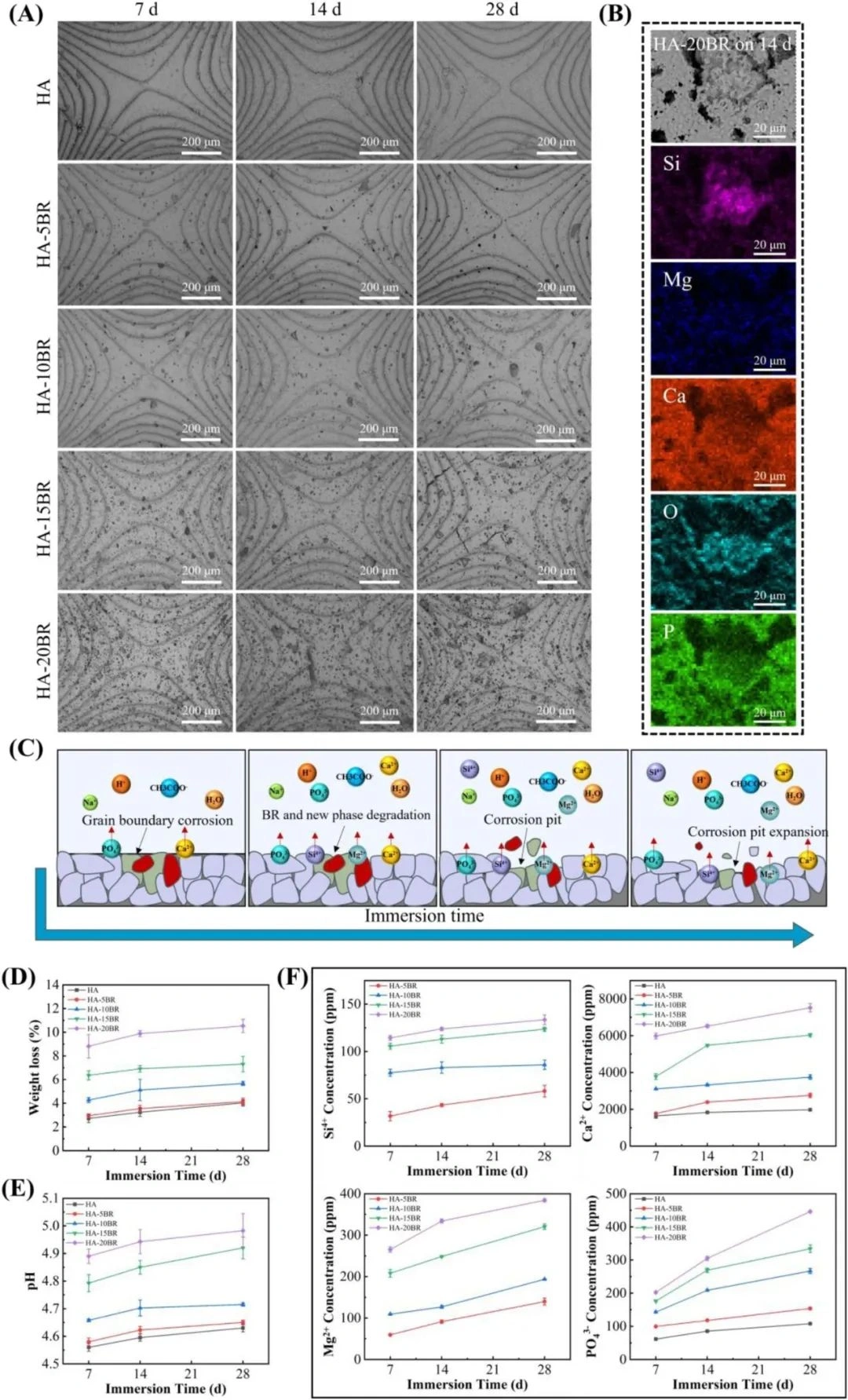
△Fig. 7, Degradation behavior of HA/BR stent. (A) Surface topography of SEM. (B) EDS mapping element analysis of corrosion pits. (C) Schematic diagram of degradation. (D) Weight loss. (E) pH change of buffer solution. (F) Ion release behavior of Ca 2+, Mg 2+, Si 4+ and PO 43-.
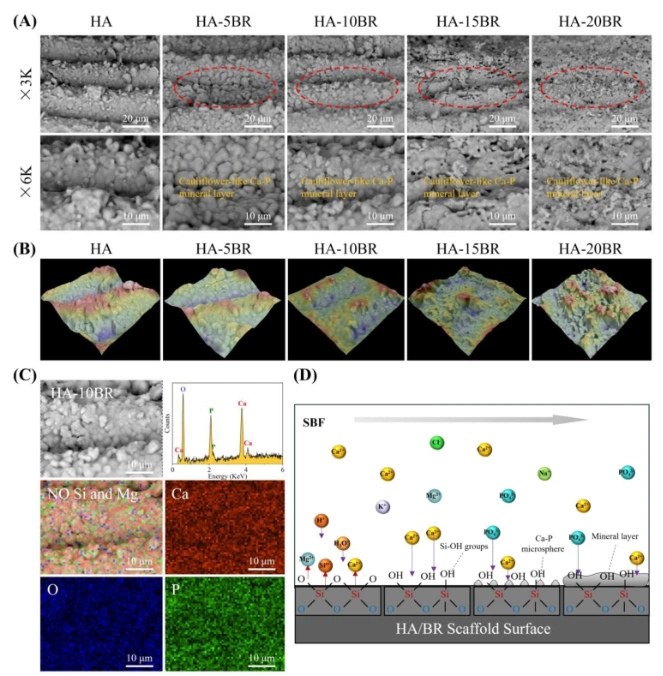
△Fig.8, The effect of BR content on biomineralization of HA/BR stents was obtained through SBF immersion test for 3 days. (A) Surface morphology analysis was performed by SEM. (B) 3D roughness reconstruction image. (C) Surface element analysis through EDS mapping. (D) Schematic diagram illustrating the biomineralization process, namely the formation and growth of calcium phosphate deposits on the surface of the scaffold.
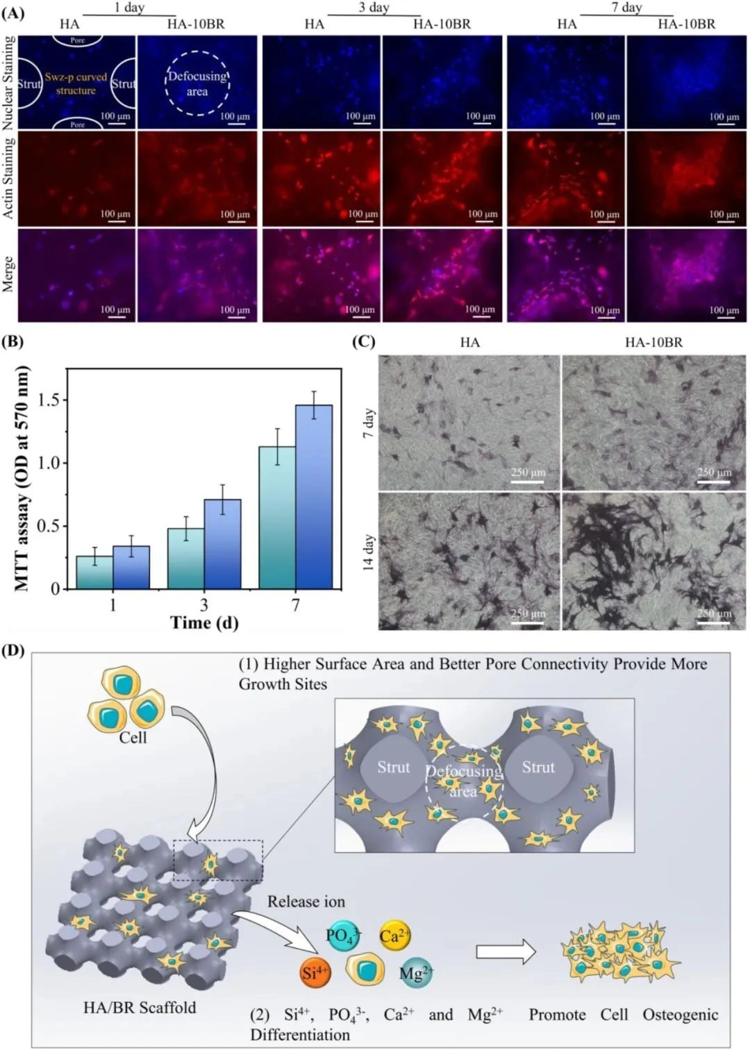
△Fig. 9, the proliferation and osteogenic differentiation behavior of MC3T3 cells were observed by fluorescence staining and ALP staining. (A) Nuclear/actin staining with rhodamine-labeled phalloidin and Hoechst 33342. (B) MTT determination. (C) ALP staining. (D) Schematic diagram of HA/BR scaffolding promoting cell growth.
Conclusion
This study proposed a method to enhance the performance of hydroxyapatite (HA) bone scaffolds by ion substitution strategy. The scaffolds with TPMS porous structure were prepared by desktop DLP technology. The scaffold surface was smooth and had no obvious defects.
During the sintering process, extending the holding time can improve the degree of densification and increase the grain size. The 2-hour holding time achieved the best balance between compressive strength and modulus. The brinstone doping generated a second phase through ion substitution, inhibited grain growth and increased microporosity. With the increase of BR doping, the microporosity increased, and the compressive strength and modulus decreased. Rapid ion release promoted mineral nucleation and growth, and significantly enhanced the biomineralization performance after 3 days of culture in simulated body fluid (SBF). HA-BR scaffolds showed excellent cell proliferation and osteogenic differentiation ability due to their porous structure and enhanced ion release.