
Industry Insights
Recently, the team led by Eoin D. O'Cearbhaill at the University College Dublin's Biomedical Engineering Center published a study in "Applied Materials Today" titled "Low-cost fabrication of digital light processing 3D printed conical microneedles for biomedical applications," utilizingDigital Light Processing (DLP) 3D printingManufacturingLow costConical microneedlesa new method.
Research Content
In this issue, let's talk about
some new advancements in microneedle technology in the medical field.
Traditional
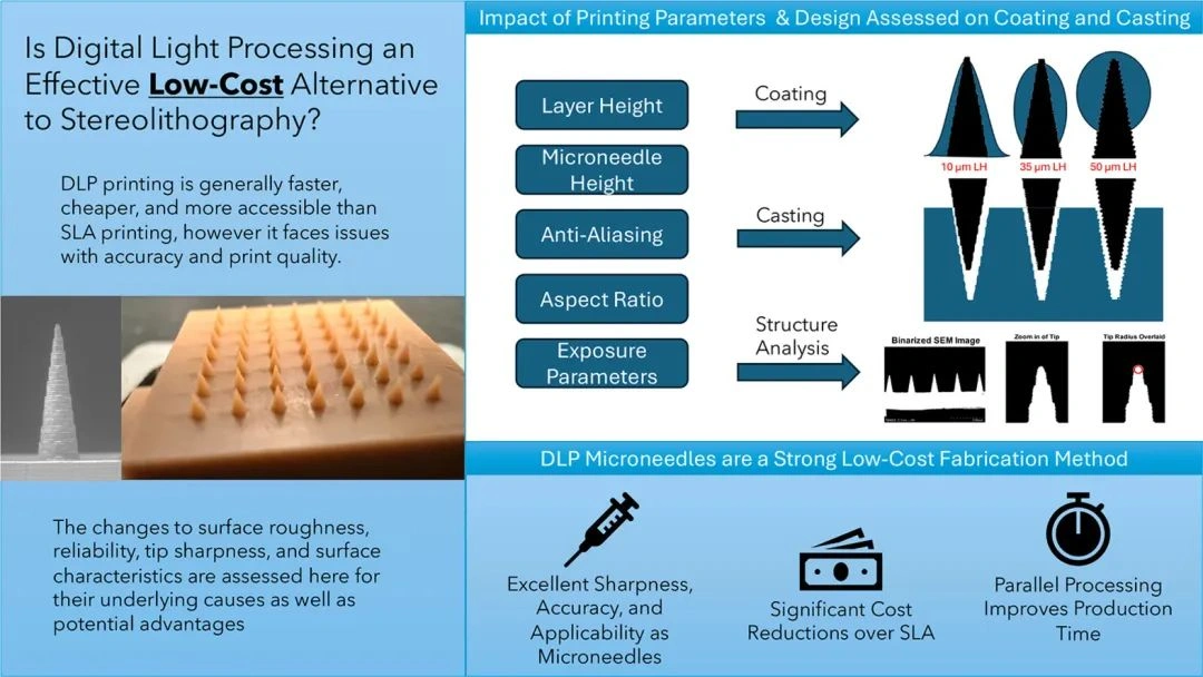
injectionmethods are feared by many because they can be painful! They may also lead to infections! Moreover, they are not very convenient to perform. However,microneedlesare different; they are small and almost painless during insertion, making them increasingly popular. Especially sincethey have a stronger ability to penetrate the skin and deliver medication more effectively. However, the traditional manufacturing methods for microneedlesare costly, complex, and not easy for mass production.Among existing 3D printing technologies, DLP technology excels in precision and surface quality, making it particularly suitable for manufacturing structures that are fine to the micron level. This article proposes a new solution, exploring the use of DLP 3D printing toConical microneedlesachieve easier operation, lower costs, higher precision, and faster speedsfor manufacturing conical microneedles.
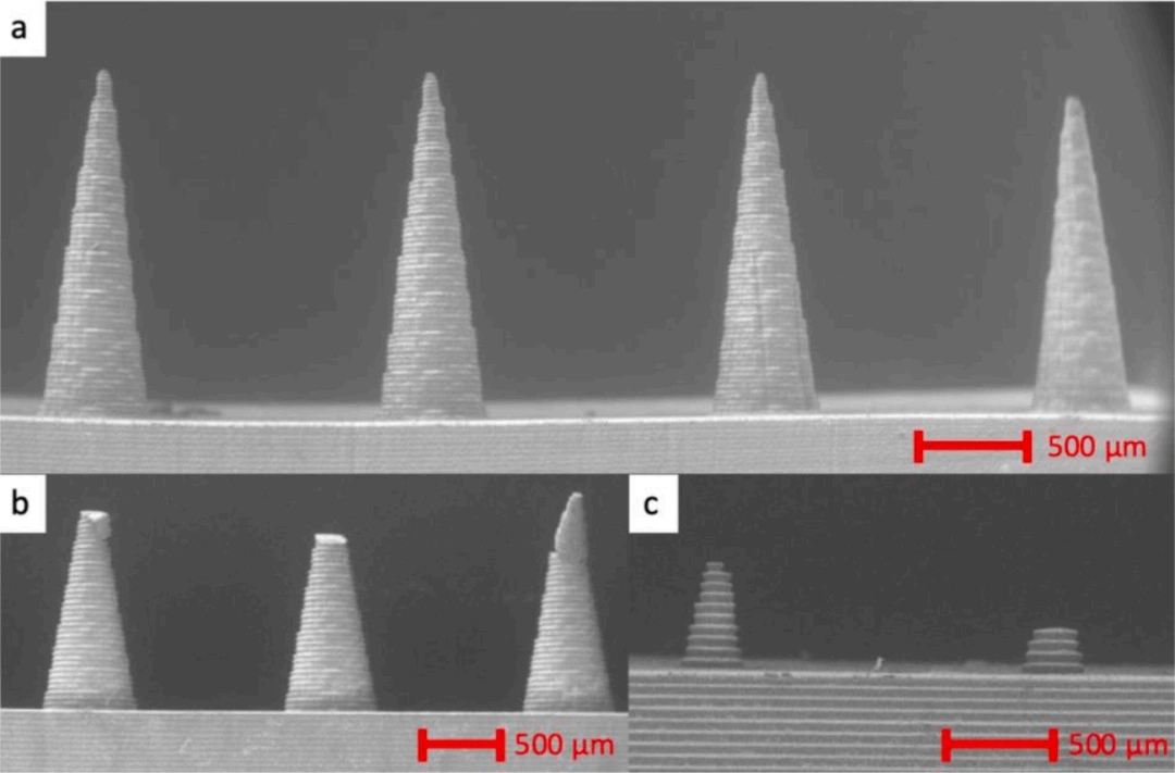
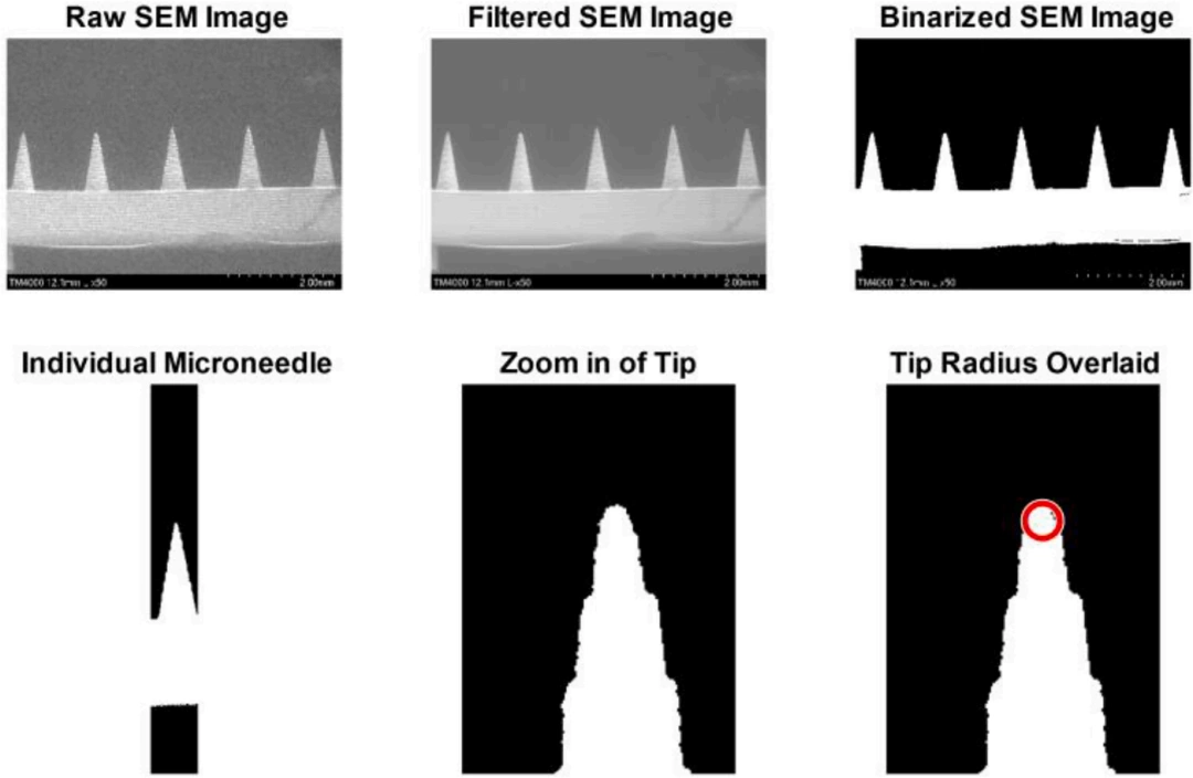
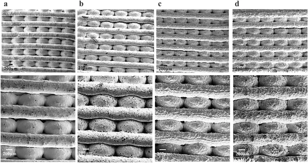
The following are the research methods and data from the article:△Figure 1, a schematic diagram of the conical structure and the effects of centered and non-centered structures within pixel boundaries during DLP printing.△Figure 2, a) A row of neatly printed DLP microneedles showing the expected shape. b) Different modes of microneedle breakage. c) 0.8 mm microneedles - intact (left), broken or malformed (right).△Figure 3, extracting microneedle geometries from SEM images.
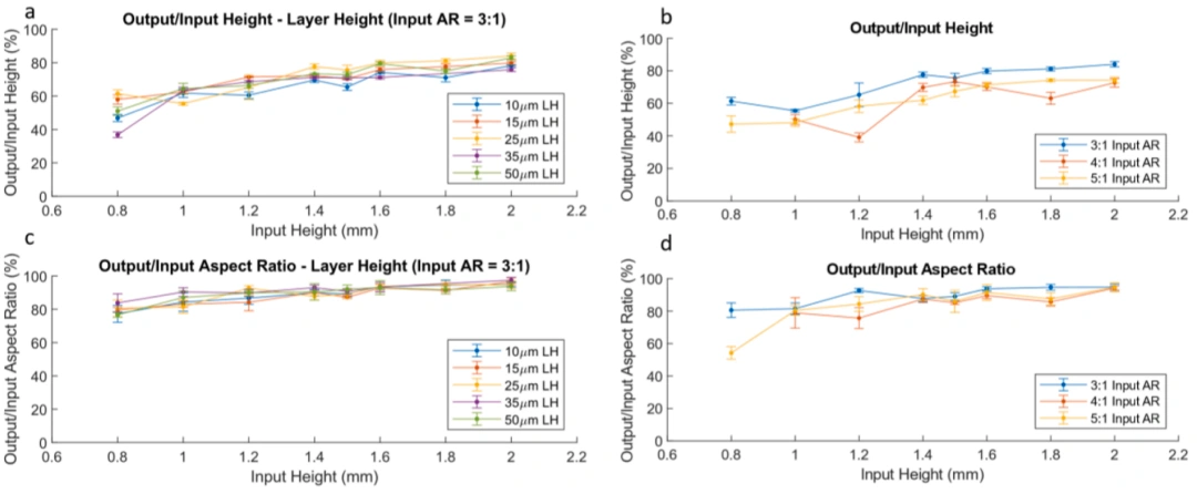
△Figure 4, the impact of needle tip input height (a) and aspect ratio (b) on output height and aspect ratio. The effect of setting print layer height on output height (c) and aspect ratio (d).

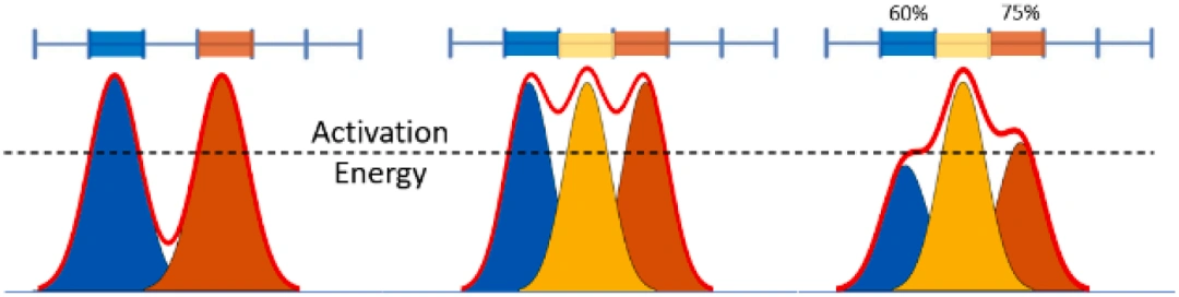
△Figure 5, estimated energy intensity levels in the photopolymer medium due to pixel alternation, complete or partial illumination. The red line indicates the total energy experienced in the medium. The blue line indicates the pixel boundaries.
△Figure 6, group comparisons of tip radii collected from the entire microneedle height range, with layer heights ranging from 10 to 50 µm. * indicates p < 0.1, *** indicates p <<< 0.01.
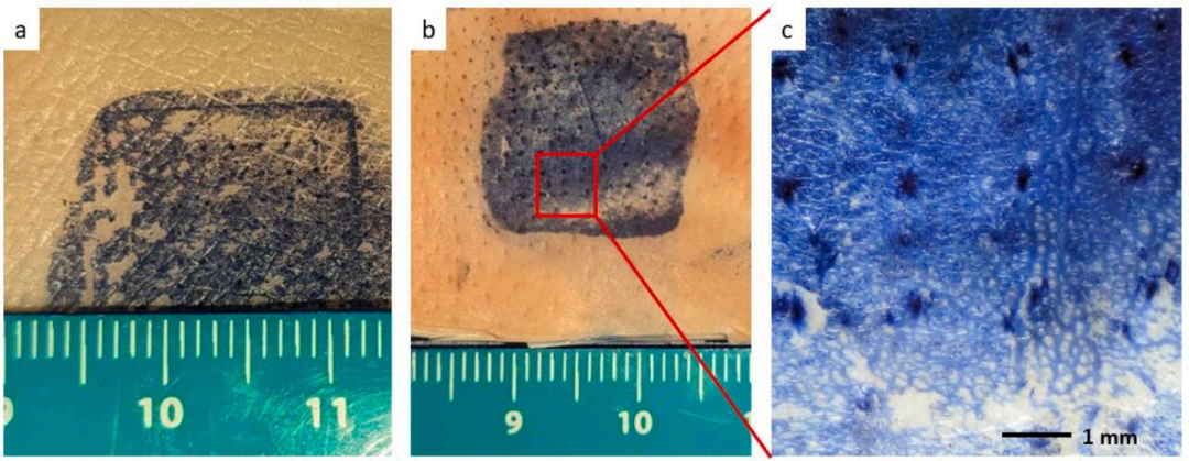
△Figure 7, results of insertion tests in pig tissue. After microneedle insertion, tissue staining application, rinsing, and wear, the results show the insertion profiles in (a) pig abdominal skin and (b) pig ear skin, along with illustrations for clarity (c).
Conclusion
If injections are no longer so painful, that is definitely good news for many people. These microneedles can achieve this by delivering medication directly to the skin surface, reducing the pain associated with traditional injections. This is a significant blessing for children who fear injections or adults who have a phobia of needles.
These microneedle arrays can not only be used for drug delivery but also serve as sensors and diagnostic tools. Because microneedles are fine enough to directly extract blood or other bodily fluids for analysis, rapid diagnosis can be performed.
Overall, manufacturing conical microneedles using DLP 3D printing technology is not only cost-effective but also holds great potential for biomedical applications. Research shows that
these microneedles meet precision standards and have enormous application prospects in drug delivery, painless injections, and vaccination.
With the continuous advancement of 3D printing technology, the manufacturing costs of these microneedles may decrease further in the future, which will help their widespread clinical application.
These microneedle arrays can not only be used to deliver drugs, they can also be used as sensors and diagnostic tools. Because the microneedles are small enough, blood or other body fluids can be directly extracted for analysis, so that diagnosis can be made quickly.
In general, the use of DLP 3D printing technology to manufacture conical microneedles is not only low-cost, but also has great potential in biomedical applications. Studies have shown that these microneedles fully meet the accuracy standards and have great application prospects in drug delivery, painless injections, vaccinations, etc. With the continuous advancement of 3D printing technology, the manufacturing cost of these microneedles may be lower in the future, which will help them become more popular in clinical applications.