
Industry new knowledge
Recently, He Fupo's team from the School of Mechanical and Electrical Engineering of Guangdong University of Technology published a study titled Improving mechanical strength and osteo-stimulative capacity of 3D-printed magnesium phosphate ceramic scaffolds by magnesium silicate additives in Ceramics International,Improving the performance of 3D printed magnesium phosphate (MgP) ceramic scaffolds by adding magnesium silicate (MgSi).
Original link: https://doi.org/10.1016/j.ceramint. 2024.08.197
Adventure Technology official website: http://www.adventuretech.cn/
If it cannot be opened, please copy the URL to the browser.
research content
The inorganic component of bone is mainly a composite of phosphate and metal elements,Magnesium (Mg)It is the fourth most abundant metal element in bone and is essential for bone growth and repair.Magnesium phosphate (Mg (PO½) ₂)Ceramics have become a potential material for bone defect repair due to their high mechanical strength, rapid degradation rate and good bone formation ability. However, its bone stimulation ability still needs to be improved. To improve performance, the incorporation of nutrients such as strontium, lithium, and silicon has been shown to promote bone formation and improve cellular responses.Silicon (Si)It can promote bone cell activity, but the release rate is limited in phosphate ceramics.
Magnesium silicon (Mg₂ SiOH4)Because of its good biocompatibility and bioactivity, as a composite material, it can maintain a magnesium-rich microenvironment and continuously release silicon to further promote bone formation.In this study, the Mg (PO₂)/Mg₂ SiO₂ composite ceramic support was prepared by 3D technology.The physical and chemical properties, in vitro degradation and cell reaction were evaluated.
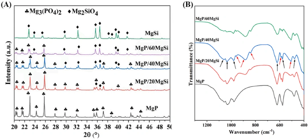
Figure 1,(A) XRD pattern of MgP/MgSi ceramic composite scaffold sintered at 1100°C. (B) The FTIR spectrum of MgP/MgSi ceramics; the black arrow indicates the absorption band of PO-4, and the red arrow indicates the absorption band of SiO4.
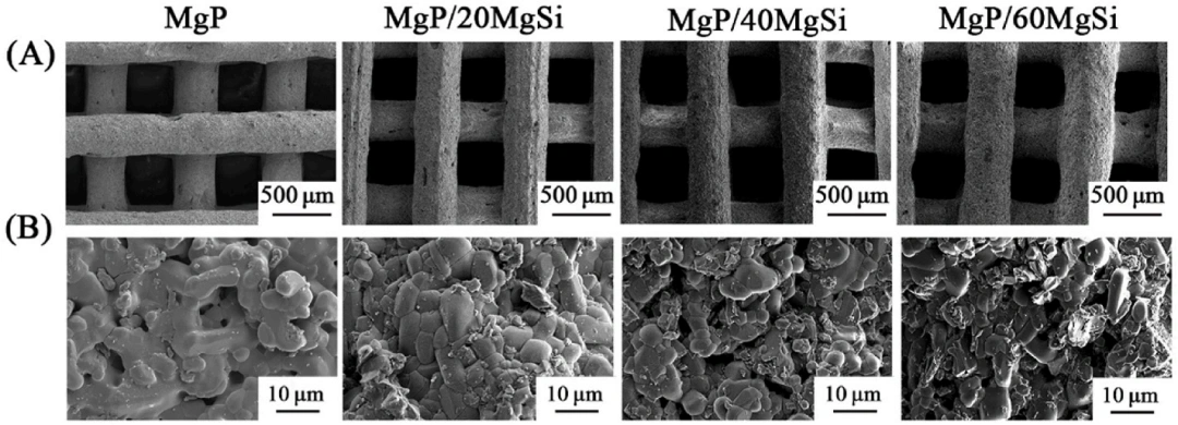
2, SEM images of 3D printed MgP/MgSi ceramic scaffolds:(A) image of macroporous morphology,(B) image of microstructure.
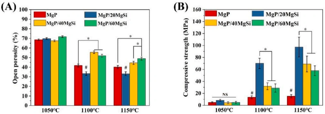
△ Figure 3, Porosity (A) and compressive strength (B) of MgP/MgSi scaffolds (N = 6,* p < 0.05,#p <0.05). * indicates significant difference between two groups,# indicates significant difference compared to all other groups, NS indicates no significant difference between groups.
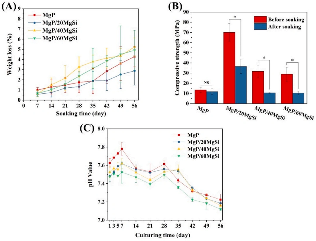
4, Weight loss (A), change in compressive strength (B) and change in pH (C) of MgP/MgSi scaffolds after immersion in Tris-HCl solution (N = 5,* p <0.05). * indicates significant difference between two groups, NS indicates no significant difference between groups.
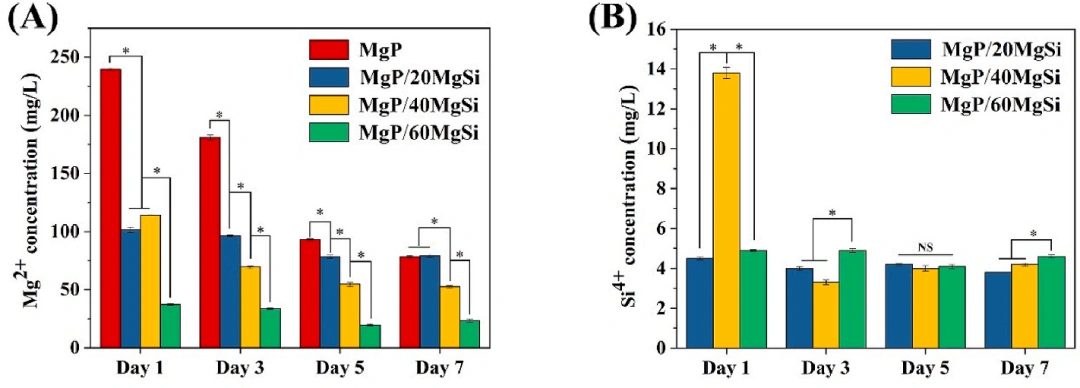
△ Figure 5, Mg² concentration (A) and Si² concentration (B) released into the medium from the scaffold (N = 3,* p <0.05). * indicates significant difference between two groups, NS indicates no significant difference between groups.
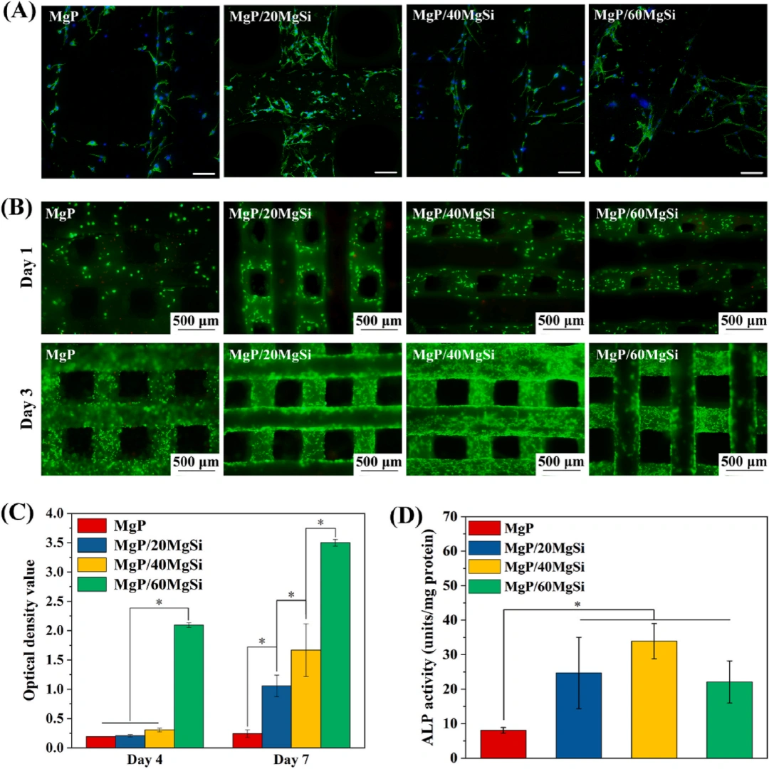
6,(A) Confocal images of the cytoskeleton of mBMSCs attached to the scaffold. Scale bar = 100 μm. (B) Fluorescent images of viable and dead cells on scaffolds. (C) Proliferation activity of mBMSCs on scaffolds. (D) Quantitative ALP activity of mBMSCs co-cultured with ceramic scaffolds (N = 4,* p <0.05). * indicates a significant difference between the two groups.
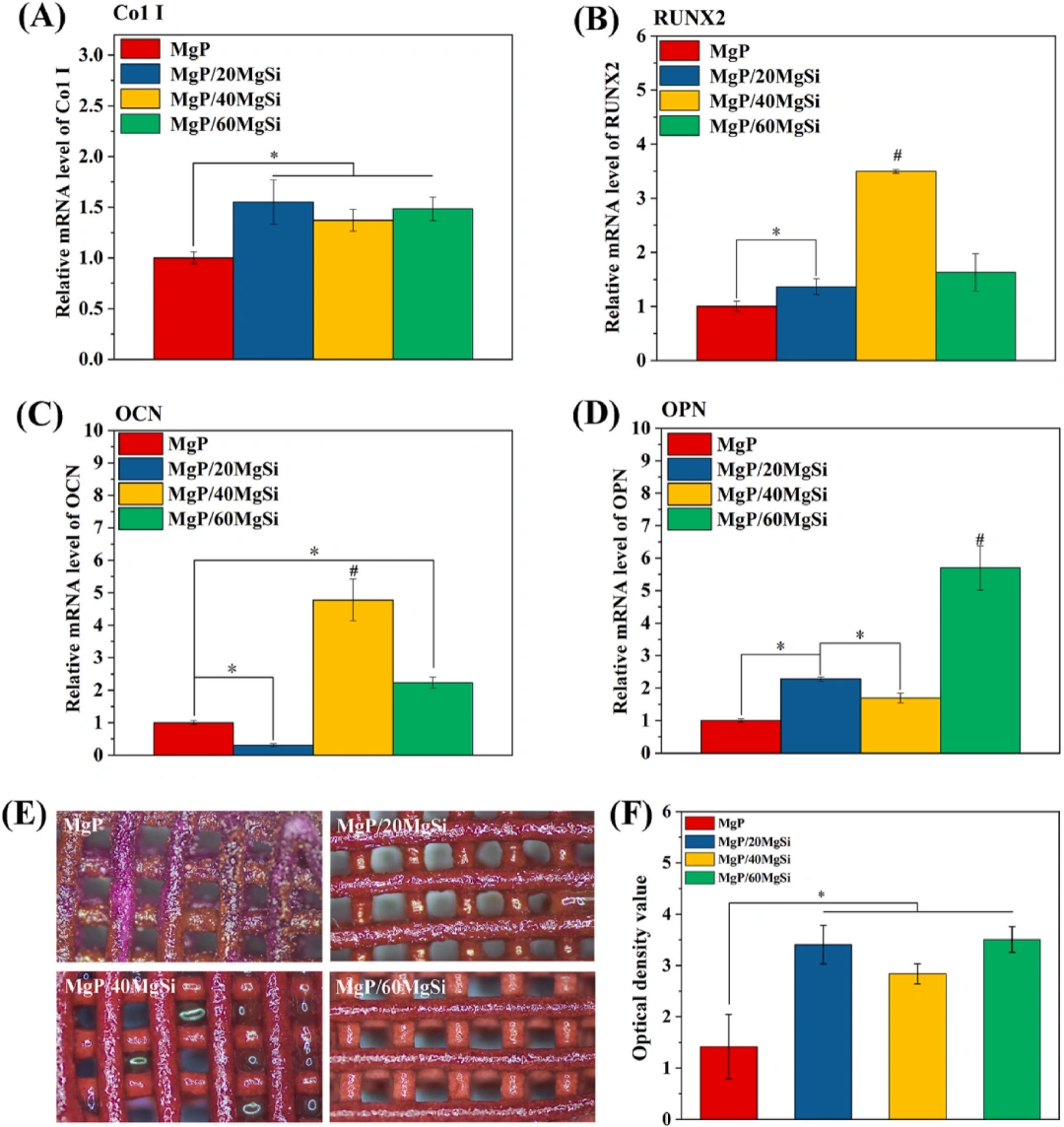
7,(A-D) Osteogenesis-related gene expression of mBMSCs cultured on MgP and MgP/MgSi scaffolds (A: Col I;B: RUNX2;C: OCN;D: OPN). (E) A photograph of ARS staining of mBMSCs after 14 days of culture on scaffolds. (F) Quantification of calcium mineral formation after 14 days of incubation of mBMSCs on scaffolds (N = 4,* p < 0.05,#p <0.05). * indicates significant difference between two groups,# indicates significant difference compared to all other groups.
Conclusion
MgSi did not react with MgP during sintering of MgP/MgSi ceramic composite scaffolds. The addition of MgSi results in a significantly higher compressive strength of the MgP/MgSi scaffold. The MgP/MgSi scaffold gradually degrades in aqueous solution, resulting in a decrease in mechanical strength. Mg and Si ions with bone stimulating effect released from MgP/MgSi scaffolds promoted the growth of mBMSCs and the expression of osteogenesis-related genes.
In general, MgP/40MgSi and MgP/60MgSi scaffolds have high porosity, mechanical strength and strong bone stimulation, which are potential biomaterials and are expected to effectively repair bone defects.