
A denture is two materials with one weak joint. The artificial teeth need hardness and wear resistance; the denture base needs toughness and resilience. In a conventional DLP workflow the two are printed separately, cleaned, then joined with a light-cured adhesive — and that bonded junction becomes the part most likely to fail in service.
A study published in Dental Materials (2026) asked whether a dual-vat DLP printer can remove that joint altogether. The authors, from Guangzhou Medical University with a co-author from Shenzhen Adventure Technology, printed denture teeth and denture base in a single multi-material build and compared it with the standard print-then-bond route, using the same ISO test methods applied to conventional denture polymers.
The short answer: the tooth–base interface became more than twice as strong, fracture behaviour became more consistent, and the material showed no cytotoxicity. Flexural strength, however, did not rise above the bonded group — and that detail is worth reading carefully.
1. Four groups, one variable
All specimens were printed on ADT DLP hardware; the multi-material group used a dual-resin-tank system. The reported print parameters were 1920 × 1080 projection, 405 nm, and 50 µm layers. Four groups isolated the effect of the printing strategy:
- PT — artificial denture teeth resin printed alone: PhEMA and UDMA with 20–30% SiO₂ filler.
- PB — denture base resin printed alone: Bis-EMA, HDDMA and 1–5% HEMA, with no filler.
- PT-PB — the two resins printed separately, then bonded with unpolymerized base resin and light-cured for 10 minutes, mimicking the conventional assembly step.
- MPT-B — teeth and base co-printed in one build on a dual-resin-tank multi-material DLP printer, with no adhesive layer.
Test specimens were disks (Ø10 or Ø14 mm × 2 mm), plus a tooth-sized disk joined to a base disk for shear testing. Water sorption and solubility followed ISO 20795-1, biaxial flexural strength followed ISO 6872, surface texture was measured to ISO 25178-2, and cytotoxicity followed ISO 10993-5/-12.
2. How the two-vat build works
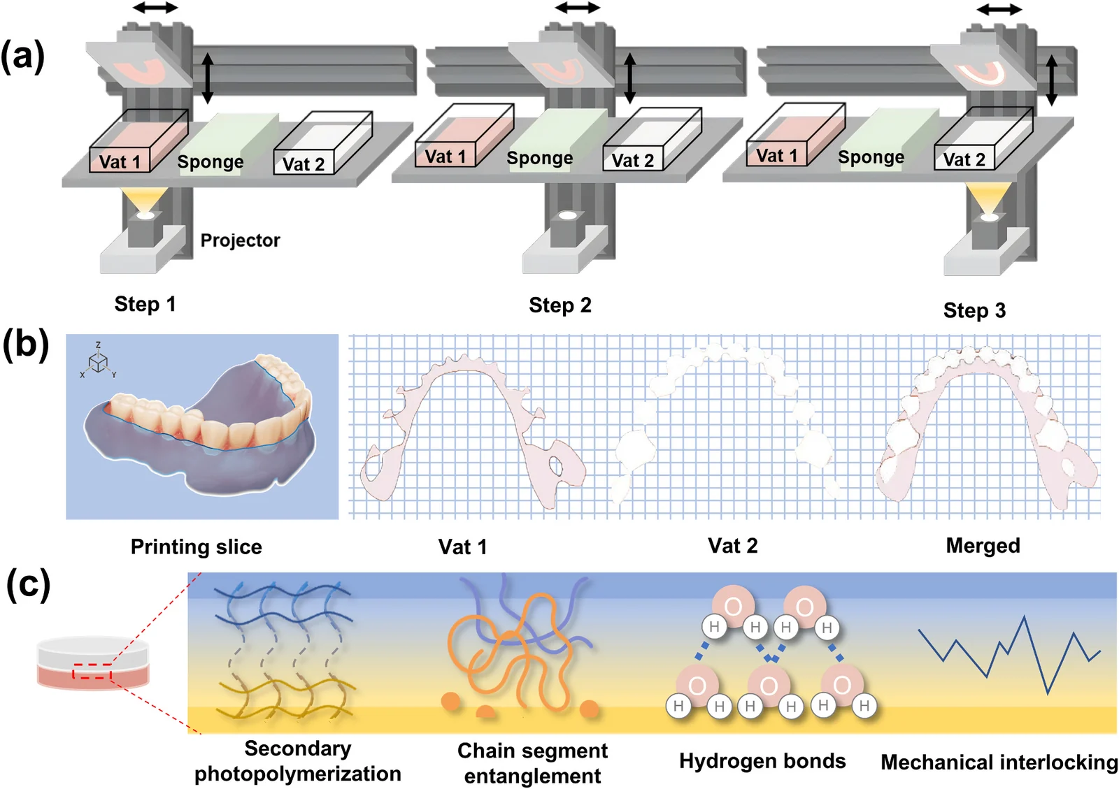
The machine carries two resin vats. The build platform prints the base region in the first vat, passes a sponge station that removes excess slurry, then prints the tooth region in the second vat — completing one layer. If a layer contains only one material, the platform stays in that vat and the cleaning step is skipped. The cycle repeats layer by layer, so the two materials meet inside the part instead of being glued afterwards.
Why the junction holds is not fully settled. The authors attribute it to a combination of secondary photopolymerization, interdiffusion and chain-segment entanglement, hydrogen bonding, and mechanical interlocking from the interfacial topography. They did not quantify these interactions directly and describe the mechanism as hypothesis-generating — a fair caveat, and one worth keeping when the result is quoted.
3. The joint: 7.96 MPa becomes 17.78 MPa
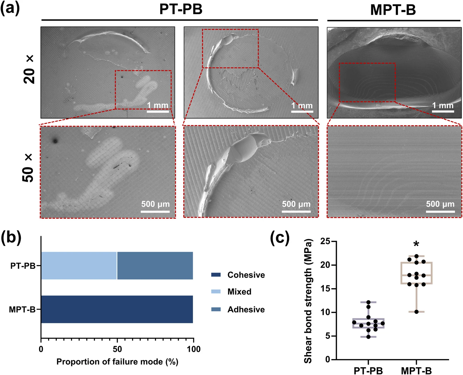
Shear bond strength was 17.78 ± 3.22 MPa for the multi-material group against 7.96 ± 2.03 MPa for the bonded group (p < 0.0001) — a factor of 2.2.
The failure modes explain more than the number does. The bonded group failed 50% adhesively and 50% mixed: the crack ran along the adhesive interface, which is exactly where a clinical denture would debond. The multi-material group failed cohesively in every specimen — the joint held, and the crack was forced through the base resin instead. In other words, the interface stopped being the weakest link. For context, the paper cites shear bond values for CAD/CAM denture systems between roughly 3.4 and 18.1 MPa depending on surface treatment; the multi-material result sits at the top of that range.
4. Surface: same DLP texture, lower micro-complexity
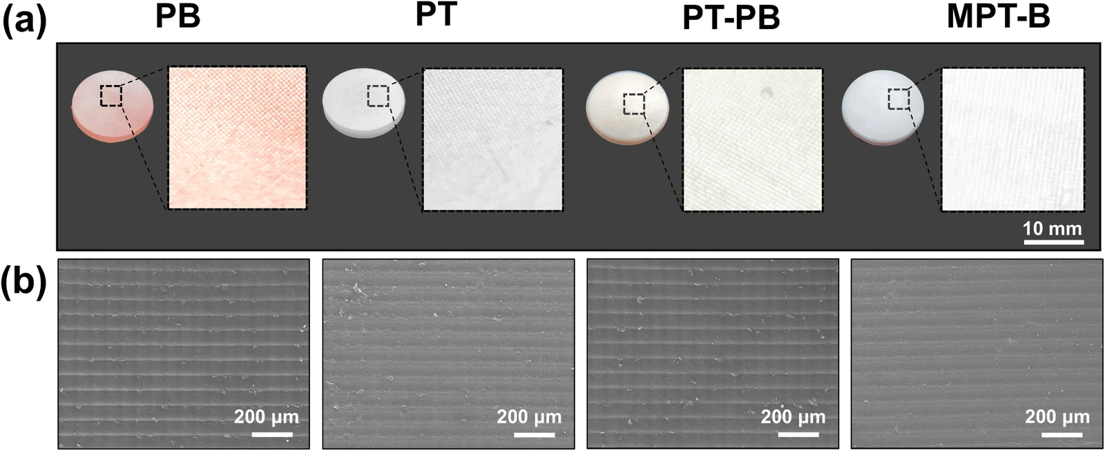
Every group showed the pixel-grid texture typical of DLP at a 0° build angle. The base resin alone was the roughest: Sa 2.47 ± 0.61 µm and Sz 16.09 ± 3.51 µm. The multi-material group measured Sa 1.33 ± 0.43 µm — significantly smoother than the base-only group (p = 0.0151) — and had the lowest Sdq and Sdr of all four groups: the lowest local slope and the least added surface area from micro-texture.
The authors are careful about what this means: lower roughness and lower micro-complexity suggest easier cleaning and better esthetic stability, but they do not prove less biofilm formation. Microbial adhesion on denture polymers depends on surface chemistry and material properties as well as topography. Treat the roughness result as a cleanability indicator, not a clinical claim.
5. Water: the bonded interface takes in more water
Water sorption was 5.86 ± 0.68 µg/mm³ for the multi-material group versus 9.81 ± 2.99 µg/mm³ for the bonded group (p = 0.0163). The base-only group measured 5.96 ± 0.77 µg/mm³. All groups stayed below the ISO 20795-1 limit of 32 µg/mm³.
Solubility is the more revealing figure. The bonded group was the only one with a positive value (+0.78 ± 0.37 µg/mm³), meaning net mass loss. The base-only, teeth-only and multi-material groups were negative (−2.95, −4.93 and −4.07 µg/mm³), which indicates water uptake exceeding the mass lost to leaching — a known behaviour of light-cured resin materials. All groups still conformed to the ISO 20795-1 limit of 1.6 µg/mm³.
The explanation the authors give is process-level: the bonded group's adhesive layer was cured through two polymers, so light was attenuated by absorption and scattering, and conversion at the interface may be incomplete. The base resin also contains 1–5% HEMA, a hydrophilic monomer. A discrete, under-cured interface gives water and unreacted monomer a path that a continuous printed junction does not have.
6. Flexural strength: a tie, and that matters
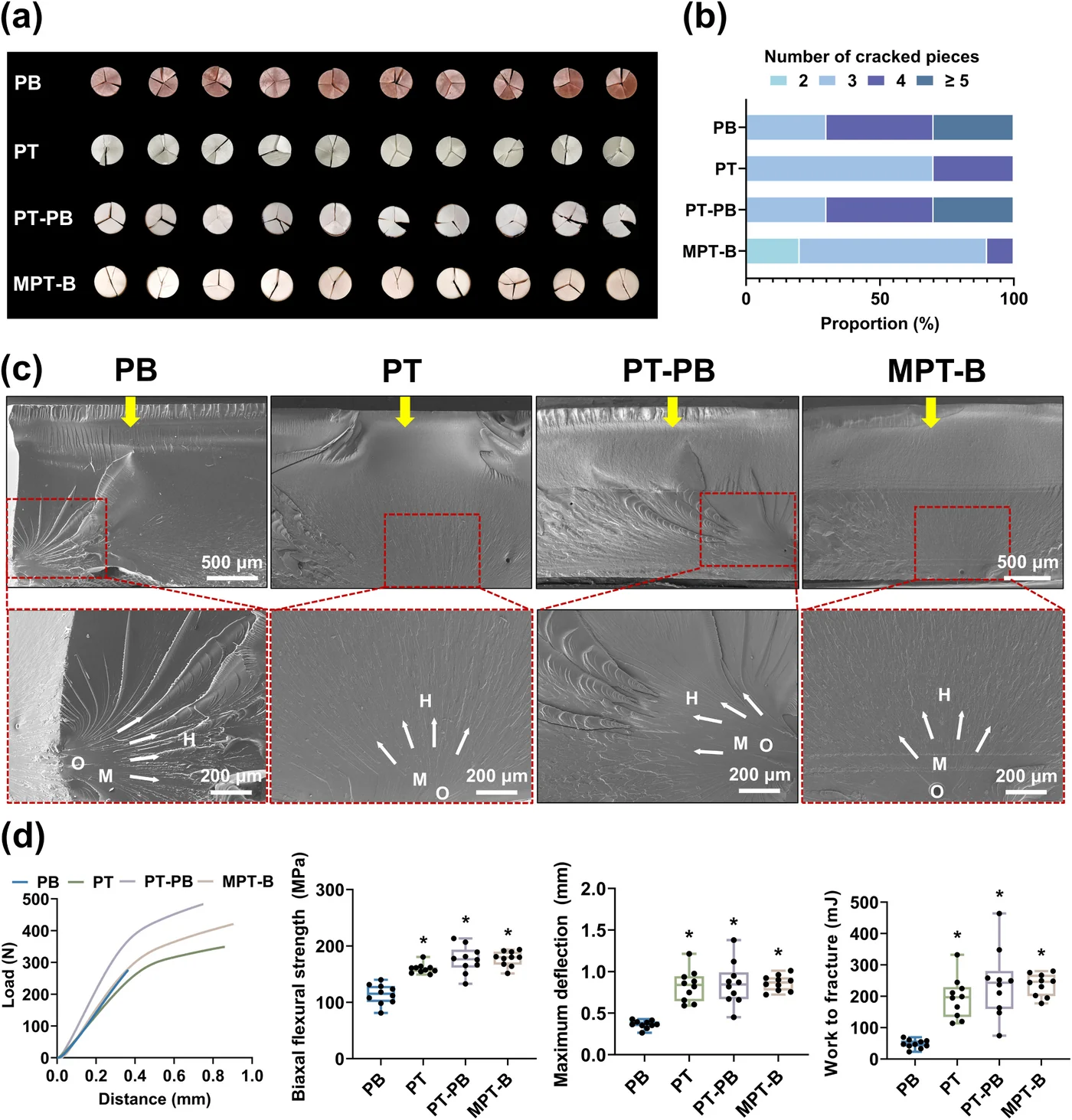
The base-only group was the weakest at 114.2 ± 17.7 MPa. The teeth-only, bonded and multi-material groups were all significantly stronger than that, and — this is the part to read closely — there was no significant difference among the three (p > 0.05). The paper's conclusion says flexural strength was higher for multi-material specimens than for conventionally bonded ones; the reported statistics show no significant difference between those two groups. The accurate summary is: dropping the bonding step did not cost flexural strength.
What did change was consistency. Every multi-material specimen broke into exactly three fragments, while the bonded and base-only groups produced four or more pieces in several specimens. The bonded group also showed wider scatter in strength and displacement than the multi-material group. Fractography showed a smoother junction with fewer secondary cracks in the multi-material group, and a more tortuous, crack-branching interface in the bonded group. For a production process, predictable fracture behaviour is worth more than a marginal mean.
7. Cytocompatibility: no signal
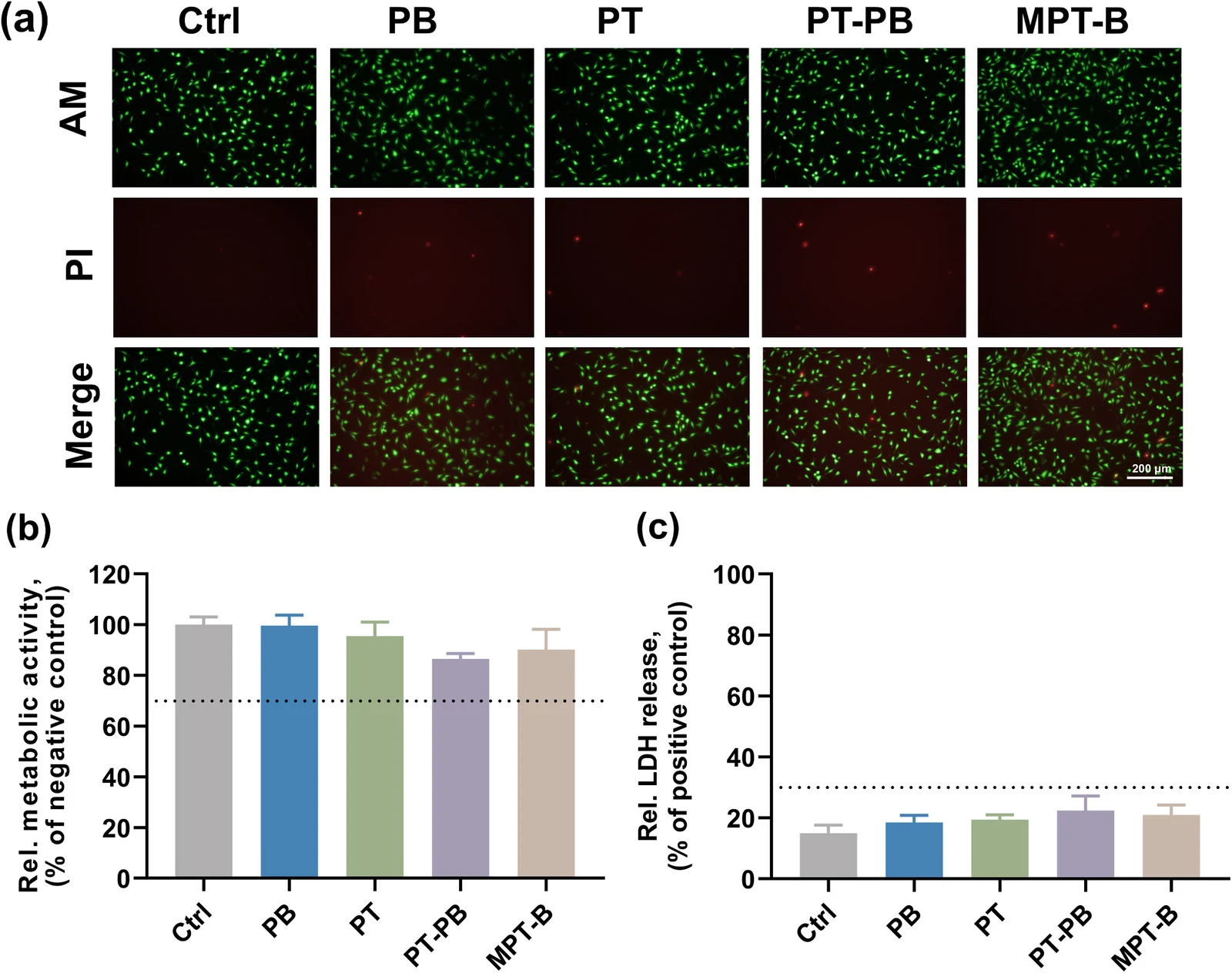
Extracts from all four groups were tested on L929 mouse fibroblasts for 24 hours. Metabolic activity stayed above 70% of the negative control in every group, and LDH release stayed below 30% of the positive control — both inside the ISO 10993-5 non-toxic range. Live/dead staining showed intact membranes and normal cell morphology.
The caveat: the study screened cytotoxicity but did not quantify residual monomers by analytical chemistry. Passing a screening test is not the same as a full leachables profile.
8. What the study does not show
- Short-term aging only. Water sorption and solubility were measured after 7 days per ISO 20795-1 — a standardized baseline, not years in the mouth. No thermocycling and no cyclic mechanical loading.
- No milled or heat-polymerized control. The comparison is between DLP workflows, so it does not position multi-material printing against established denture fabrication routes.
- Disk specimens, not complete dentures. The slicing example in Figure 1 shows a full denture, but the mechanical tests used disks and a tooth-sized bonded pair.
- No residual-monomer analysis, no time-and-cost study, no wear or fatigue data — all named by the authors as future work.
- One resin pair from one manufacturer. The results are material-specific; another teeth/base combination may behave differently.
9. What it means for digital denture production
The value here is not a stronger plastic. It is the removal of a process step whose quality is hard to see. A bonded denture can look perfect and still carry an under-cured, HEMA-containing layer exactly where the tooth meets the base. Printing that junction instead of gluing it removes the hidden variable — and the data show the junction moving from "the likely failure point" to "stronger than the base resin".
For labs and manufacturers, the practical question is whether the workflow fits: a dual-vat machine, two resins, and a slicing step that assigns material region by region. ADT's Multi-Material DLP 3D Printer is built for exactly this — different materials between layers and within the same layer, with automatic sponge cleaning for high-viscosity slurries.
This was an in vitro study. It does not certify a denture product, and it does not replace clinical evaluation. What it does provide is measured interface data under the same ISO methods used for conventional denture polymers — the kind of evidence that lets a lab decide whether the multi-material route is worth building a workflow around.
Source: Wu K, Zhang K, Yan Q, Zhu C, Huang J, Zhu P, Wang F, Li P. Multi-material DLP printing for denture materials: investigation of surface characteristics, physico-mechanical properties, and cytocompatibility. Dental Materials (2026). doi:10.1016/j.dental.2026.02.021. Figures reproduced from the cited article for discussion.