
>25 % of the patients who receive orthopedic implants have been reported with implant-associated osteomyelitis, which can result in inflammation, osteolysis, and aseptic loosening of implants. Current treatment methods doesn't ensure defect healing and prevention from reinfection. Thermoplastic-based 3D-printed scaffolds offer a bioresorbable, biocompatible, and mechanical strong implant system. However, the hydrophobicity and bio-inertness of these polymers prevent their use in clinics.
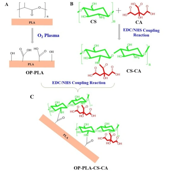
Fig. 2. Schematic diagram showing surface modification strategy of 3D printed PLA. (A) Oxygen plasma of PLA scaffolds (B) Synthesis of chitosan –citric acid (CS-CA) via EDC/NHS coupling reaction. (C) covalent functionalization of CS-CA into the surface of the 3D-printed scaffold.
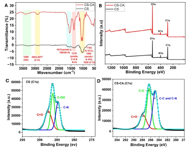
Fig. 3. (A) FTIR spectra of chitosan (CS) and citric linked chitosan (CS-CA). Characteristics peaks of CS were obtained at 2855 cm−1, 1544 cm−1, and a broad peak from 3653 to 3000 cm−1, corresponding to CH stretching, the vibration of NH, and the vibration of OH, respectively. FTIR of CS-CA showed an increased intensity and broadening of the OH peak and increased peak intensity at 1655 cm−1 and 1544 cm−1 indicating the formation of new amide bonds. (B) XPS spectra and elemental analysis for CS and CS-CA demonstrated the increased intensity of the oxygen element after citric linkage with chitosan. High-resolution XPS spectra of (C) CS for C1s, and (D) CS-CA for C1s, along with deconvolution curves.
In this study, we developed dual functionalized scaffolds with osteogenic and antibacterial properties by immobilizing citric acid-linked chitosan on oxygen plasma etched 3D-printed PLA scaffolds through an EDC-NHS coupling reaction. Acellular mineralization of these scaffolds in DMEM demonstrated the deposition of crystalline hydroxyapatite.
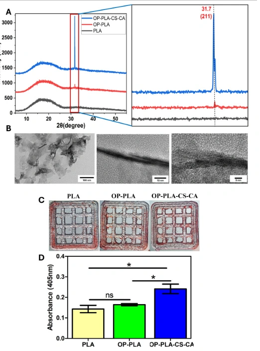
Fig. 8. (A) XRD spectra demonstrated the nature of the deposited mineral on 3D-printed PLA scaffolds where surface-modified scaffolds showed a peak at 31.7 for 211 planes, indicating the formation of hydroxyapatite crystals. A highly intense peak was found in OP-PLA-CS-CA scaffolds than OP-PLA whereas no peak was observed in PLA scaffolds, indicating the deposition of amorphous phase CaP. (B) TEM micrographs showed long elongated needle-like HA crystals mechanically removed from OP-PLA-CS-CA scaffolds. Qualitative and quantitative analysis of mineral deposits after acellular mineralization using alizarin red staining. (C) Optical images of stained samples demonstrating complete surface staining in the CS-CA coated scaffolds compared to OP-PLA and PLA scaffolds. (D) Quantitative analysis of mineral content revealed the highest levels in CS-CA coated scaffolds, while PLA and OP-PLA scaffolds exhibited no significant differences. (n = 3, *p < 0.05 PLA and OP-PLA-CS-CA, and OP-PLA and OP-PLA-CS-CA).
In addition, the antibacterial properties of these surface-modified scaffolds have been determined against E. coli and S. aureus, where the citric-linked chitosan biofunctionalized 3D-printed PLA scaffolds showed significantly higher antibacterial activity in comparison to oxygen-etched PLA and PLA scaffolds due to the synergistic effect of citric acid and chitosan functionalities.
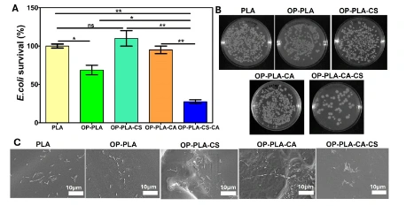
Fig. 9. Antibacterial activity of surface modified 3D-printed PLA scaffolds against E.coli bacteria (A) Antibacterial activity determined quantitatively in terms of E.coli survival % where OP-PLA-CS-CA scaffold showed the highest antibacterial activity with 70 % reduction in bacterial count (n = 3, *p < 0.05 between PLA and OP-PLA, between OP-PLA and OP-PLA-CS-CA, **p < 0.01 between PLA and OP-PLA-CS-CA, between OP-PLA-CS and OP-PLA-CS-CA and between OP-PLA-CA and OP-PLA-CS-CA.). (B) Digital images of CFU plates of E. coli treated with 3D-printed PLA (a), OP-PLA (b), OP-PLA-CS (c), OP-PLA-CA (d), and OP-PLA-CS-CA (e) scaffolds for 24 h. (C) SEM images of 3D printed scaffolds treated with E. coli demonstrated least bacterial numbers on the CS-CA coated PLA scaffolds.
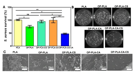
Fig. 10. Antibacterial activity of surface-modified 3D-printed PLA scaffolds against S. aureus bacteria (A) Antibacterial activity determined quantitatively in terms of S. aureus survival % where OP-PLA-CS-CA scaffold showed the highest antibacterial activity with a 50 % reduction in bacterial count (n = 3, **p < 0.01 OP-PLA and OP-PLA-CS-CA, ***p < 0.001 between PLA and OP-PLA, and ****p < 0.0001 between PLA and OP-PLA-CS-CA). (B) Digital images of CFU plates of S. aureus treated with 3D-printed PLA (a), OP-PLA (b), and OP-PLA-CS-CA (c) scaffolds for 24 h. (C) SEM images of 3D-printed scaffolds treated with S. aureus, demonstrated least bacterial numbers on the CS-CA coated PLA scaffolds.
MG-63 cells exhibited increased proliferation and osteogenic activity on the modified scaffolds compared to the PLA and OP-PLA. These 3D-printed scaffolds, coated with citric-linked chitosan, can be a potential solution to orthopedic complications such as critical-sized bone defects and implant-associated osteomyelitis.
Conclusion
The CS-CA functionalized 3D-printed PLA scaffolds have the potential to regenerate bone and prevent bacterial infection at the defect site. These scaffolds can be potentially translated into clinics, for the treatment of critical-sized bone defects while curbing implant-associated infections.
Article Source:https://doi.org/10.1016/j.ijbiomac.2024.136968